
Page 142 - 2019_03-Haematologica-web
P. 142
F. Koczian et al.
A
B
CD
EF
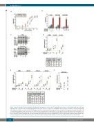
Figure 1. Chemosensitization of acute leukemia cells with PS89. (A,B) Apoptosis of Jurkat, HL-60, CCRF-CEM and vincristine resistant (VCR-R) CEM cells treated with cytostatics (ETO: etoposide, VCR: vincristine, DNR: daunorubicin) in the presence or absence of 25 mM PS89. The percentage of apoptotic cells was determined by FACS analysis after 48 h. (C) Jurkat and VCR-R CEM were cultured for 48 h in drug-supplemented medium and apoptosis was analyzed by immunoblotting. (D,E) Freshly isolated peripheral blood mononuclear cells (PBMC) and acute lymphoblastic leukemia (ALL) or acute myeloid leukemia (AML) patient-derived xenograft cells were treated with PS89 and cytostatics for 48 h or 72 h, respectively. Apoptotic cells were determined by FACS analysis and specific apoptosis was calculated in rela- tion to untreated controls. Synergism was calculated using the Bliss independence model. (F) PBMC were treated for 48 h with the indicated drugs. CD34-positive cells were identified by flow cytometry using a fluorescein isothiocyanate-conjugated CD34 antibody. Cell death was analyzed by propidium iodide staining.
548
haematologica | 2019; 104(3)
