
Page 51 - Haematologica Vol. 110 - January 2025
P. 51
PERSPECTIVE ARTICLE
dolent T-LGLL may occur, individuals must be considered one by one. Further prospective studies are needed to find a proper cutoff to define those patients who have a true pre-leukemic state rather than manifesting a byproduct of a nonspecific immune response with more limited im- pact. This is in line with the diversity established between high- and low-count MBL,84 with the latter subset being associated with a negligible risk of progression.
- Bone marrow. An evaluation of the bone marrow, if per- formed, may be of help. In fact, the absence of interstitial and intrasinusoidal cytotoxic T-cell infiltrates, or interstitial cytotoxic T-cell clusters of lymphoid cells with azurophilic granules, favors the diagnosis of T-CUS.28,85
All the above are needed to bring us to the diagnosis of T-CUS. Figure 1 illustrates the clinical landscape of T chronic lymphoproliferative disorders of LGL. T-CUS stands between reactive, polyclonal or transiently clonal lymphocytosis and indolent LGLL. It represents the benign end of the broad spectrum of these disorders, but the watershed distinguish- ing indolent T-LGLL from T-CUS is nuanced, as emphasized early. We hope to find a biomarker that differentiates be- tween T-CUS and indolent T-LGLL. Certainly, the putative detection of somatic mutations can prospectively bridge this knowledge gap, enabling a more timely identification of these borderline subsets of patients.
Understanding the evolution of T-cell clones is crucial for clinical management and future research directions
When can we be confident that we are dealing with a T-CUS instead of true T-LGLL (Table 3)? We believe that the selected circumstances previously discussed45,52-56,63-66,69 should be reconsidered on a case-by-case basis taking into account the precise criteria currently in use for T-CUS/T- LGLL in the era of genomics.
G. Semenzato et al. Chronic exposure to specific antigens (infections, tumor
antigens, protein products or graft antigens) could trig- ger the initial LGL proliferation and the emergence of a clone then confers a growth advantage on the clone per- sistence and expansion of lymphocytosis. In some cases, the acquisition over time of specific genetic alterations or permissive epigenetic changes is believed to definitely establish founding clones. This leads to clonal overgrowth and suppression of hematopoiesis, undergoing malignant transformation and then resulting in the onset of LGL leu- kemia. Are these further steps mediated by mutations or epigenetic alterations that grant the affected cells a growth advantage over their counterparts, making the rogue clone become dangerous by triggering cytopenias and autoim- munity? Consistent with this hypothesis, STAT3 mutations have not been detected in persistent cytotoxic T lympho- cyte expansions following allogeneic HSCT that remained stable over the time62 nor in inflammatory myositis.86 This absence of mutations may indicate a scenario in which an equilibrium is reached between immune system control and the triggering event(s). Should mutations disrupt this equilibrium, the clone undergoes progressive expansion with detrimental effects. These findings offer novel insights into this neglected topic, underscoring the need for future prospective studies to track early steps of disease and to determine which patients, when, and under what circum- stances progress to having full-blown disease. Steps to progression are likely mediated by multiple events, which include not only mutations but also dysregulated pathways, and the influence of the microenvironment. Indeed, the absence of any LGLL phenotype in mice expressing STAT3 mutations87 suggests that additional gene mutations or deregulation of other signaling molecules or pathways30 might be involved in association with STAT3 mutations in the pathogenesis of LGLL. Taken together, these findings underscore the complexity of the disease and the need for further research to elucidate the underlying mechanisms and the discovery of molecules/pathways that may be at- tractive for immunotherapeutic approaches.
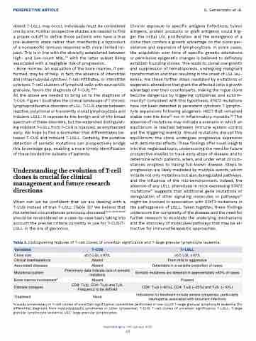
Table 3. Distinguishing features of T-cell clones of uncertain significance and T-large granular lymphocyte leukemia.
Variables
T-CUS
T-LGLL
Clone size
≤0.5 LGL x109/L
>0.5 LGL x109/L
Clinical manifestations
Absent
From mild to aggressive
Associated diseases
Absent
Detectable in a variable proportion of cases
Mutational pattern
Preliminary data indicate lack of somatic mutations
Somatic mutations are detected in approximately >50% of cases
Bone marrow involvement#
Absent
Present
Disease subtypes
CD8+ Ta/β, CD4+ Ta/β and Tg/d. Frequency to be defined
CD8+ Ta/β (~65%), CD4+ Ta/β (~25%) and Tg/d– (~10%)
Treatment
None
Indications for treatment include severe cytopenias, particularly neutropenia associated with recurrent infections
#Usually unnecessary in T-cell clones of uncertain significance; sometimes performed in low-count T-large granular lymphocyte leukemia (for differential diagnosis from myelodysplastic syndromes or other cytopenias). T-CUS: T-cell clones of uncertain significance; T-LGLL: T-large granular lymphocyte leukemia; LGL: large granular lymphocytes.
Haematologica | 110 January 2025
43
