
Page 50 - Haematologica Vol. 110 - January 2025
P. 50
PERSPECTIVE ARTICLE G. Semenzato et al.
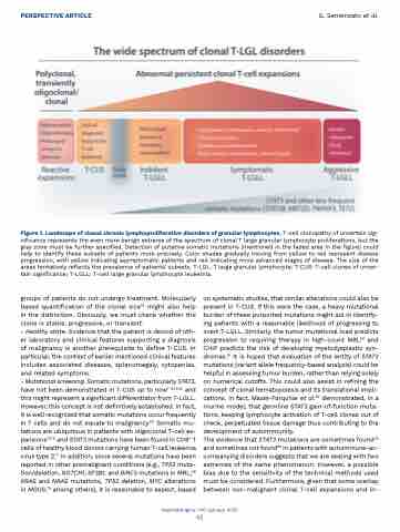
Figure 1. Landscape of clonal chronic lymphoproliferative disorders of granular lymphocytes. T-cell clonopathy of uncertain sig- nificance represents the even more benign extreme of the spectrum of clonal T large granular lymphocyte proliferations, but the gray zone must be further specified. Detection of putative somatic mutations (mentioned in the faded area in the figure) could help to identify these subsets of patients more precisely. Color shades gradually moving from yellow to red represent disease progression, with yellow indicating asymptomatic patients and red indicating more advanced stages of disease. The size of the areas tentatively reflects the prevalence of patients’ subsets. T-LGL: T large granular lymphocyte; T-CUS: T-cell clones of uncer- tain significance; T-LGLL: T-cell large granular lymphocyte leukemia.
groups of patients do not undergo treatment. Molecularly based quantification of the clonal size74 might also help in the distinction. Obviously, we must check whether the clone is stable, progressive, or transient.
- Healthy state. Evidence that the patient is devoid of oth- er laboratory and clinical features supporting a diagnosis of malignancy is another prerequisite to define T-CUS. In particular, the context of earlier mentioned clinical features includes associated diseases, splenomegaly, cytopenias, and related symptoms.
- Mutational screening. Somatic mutations, particularly STAT3, have not been demonstrated in T-CUS up to now1-3,54,62 and this might represent a significant differentiator from T-LGLL. However, this concept is not definitively established. In fact, it is well recognized that somatic mutations occur frequently in T cells and do not equate to malignancy.1,13 Somatic mu- tations are ubiquitous in patients with oligoclonal T-cell ex- pansions75,76 and STAT3 mutations have been found in CD8+ T cells of healthy blood donors carrying human T-cell leukemia virus type 2.77 In addition, since several mutations have been reported in other premalignant conditions (e.g., TP53 muta- tion/deletion, NOTCH1, SF3B1, and BIRC3 mutations in MBL;78 KRAS and NRAS mutations, TP53 deletion, MYC alterations in MGUS,79 among others), it is reasonable to expect, based
on systematic studies, that similar alterations could also be present in T-CUS. If this were the case, a heavy mutational burden of these purported mutations might aid in identify- ing patients with a reasonable likelihood of progressing to overt T-LGLL. Similarly, the tumor mutational load predicts progression to requiring therapy in high-count MBL80 and CHIP predicts the risk of developing myelodysplastic syn- dromes.81 It is hoped that evaluation of the entity of STAT3 mutations (variant allele frequency-based analysis) could be helpful in assessing tumor burden, rather than relying solely on numerical cutoffs. This could also assist in refining the concept of clonal hematopoiesis and its translational impli- cations. In fact, Masle-Farquhar et al.82 demonstrated, in a murine model, that germline STAT3 gain-of-function muta- tions, keeping lymphocyte activation of T-cell clones out of check, perpetuated tissue damage thus contributing to the development of autoimmunity.
The evidence that STAT3 mutations are sometimes found42 and sometimes not found83 in patients with autoimmune-ac- companying disorders suggests that we are dealing with two extremes of the same phenomenon. However, a possible bias due to the sensitivity of the technical methods used must be considered. Furthermore, given that some overlap between non-malignant clonal T-cell expansions and in-
Haematologica | 110 January 2025
42
