
Page 87 - Haematologica-5
P. 87
Proteomic profiling reveals complexity of AML MSC
NL-MSC (P=0.032). These findings suggest that AML- MSC tend toward senescence.
Therapy alters proteins expression in AML-MSC
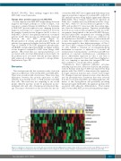
Protein expression in AML-MSC might change between diagnosis and relapse, perhaps as a result of relapse, or in response to acquired changes in AML blasts. To determine if AML-MSC protein expression was changing between diagnosis and relapse we compared expression between the samples obtained at first diagnosis (n=53) to those of AML-MSC collected from primary refractory or relapsed patients (n=54). Nine proteins are differentially expressed between the two MSC groups (Figure 5). Phosphorylated β-catenin, phosphorylated RPS6, and galectin-3 are expressed at higher levels in MSC in the sal- vage set. SMAD6, TCF4, LYN, integrin-β3, phosphorylat- ed EIF4BP1, and phosphorylated ELK1 are higher in MSC at first diagnosis compared to MSC from salvage AML patients. IPA reveals that, for canonical pathways, a set associated with osteoblast differentiation was found to be the pathway most associated with the proteins differen- tially expressed at diagnosis compared to salvage (Online Supplementary Figure S8).
Discussion
This study presents the first systematic study of protein expression differences between NL-MSC and AML-MSC. There were several notable observations. There were clear differences between the protein expression of MSC (whether from healthy donor or AML patient) and AML blast cells. This result was not surprising as one would predict different proteins would be prominent in mes- enchymal cells and cells of hematopoietic/myeloid line- age. The major observation of this research was the dis-
covery that AML-MSC have significantly different protein expression patterns compared to normal MSC, with 28 of 151 analyzed proteins being highly significantly different (FDR<0.006). These changes assumed four signatures in AML, 81% of which were very different from that of nor- mal MSC, while 6% had an identical signature to NL- MSC, and another 13% were more like the normal signa- ture than the leukemia patterns. Signature membership showed an association with cytogenetics, with 'favorable' cytogenetics being limited to the more NL-MSC-like pat- tern and 'unfavorable' cytogenetics not occurring in AML with an NL-MSC-like pattern. There was a difference in the distribution of the MSC population between men and women. The significance of these differences is not clear, but women tended to have higher percentages of Class 1 and Class 2 MSC compared to men. In leukemia progeni- tor cells, GSK3B is activated via an integrin-mediated mechanism in response to adhesion to a stromal cell exclusively in women patients.35 As ITGA2 and GSK3 are members of a protein constellation (i.e. constellation 1) that is differentially expressed in Class 1 and Class 2 (lower levels) compared to Class 3 and Class 4 (higher lev- els), it is tempting to speculate that integrin/GSK3 axis may contribute to sex-specific effects in MSC.
Furthermore, these signatures influence outcomes including response rates, remission duration and, perhaps, survival. Patients with Class 3 MSC fare much better than patients with Class 4 MSC as demonstrated by significant- ly longer remission duration and a trend toward longer OS. Changes in protein expression were often character- ized by protein-protein correlations that were reversed from those seen in normal MSC, providing insight into the nature of this dysregulation and potentially providing therapeutic targets. In NL-MSC, the signature proteins were associated with adipocyte differentiation. That normal MSC, but not AML-MSC, possess protein pathways impor-
Figure 5. A distinct set of proteins is associated with acute myeloid leukemia (AML) patient salvage status. (A) Reverse phase protein arrays (RPPA) reveals protein expression in AML mesenchymal stromal cells (MSC) differs between diagnosis and the salvage setting for 9 proteins (P=0.05; false discovery rate=0.68).
haematologica | 2018; 103(5)
819
