
Page 85 - Haematologica-5
P. 85
Proteomic profiling reveals complexity of AML MSC
are positively correlated with the protein in AML-MSC. ARC (NOL3), HSP90AA1/B1, MAPK9, PTGS2, and YWHAZ were positively correlated with β-catenin in NL-MSC but are negatively correlated with the protein in AML-MSC. Ingenuity Pathway Analysis (IPA) was performed using software on the proteins identified as differentially correlated with β-catenin in the MSC sets and β-catenin itself (i.e. CTNNB1; BCL2; BCL2L11; FOXO1; FOXO3; KIT; ITGAL; PSMD9; RPS6KB1; SFN; SRC; NOL3; HSP90AA1; HSP90B1; MAPK9; PTGS2; YWHAZ). IPA revealed these proteins were highly asso- ciated with PI3K/AKT signaling (top canonical pathway; P=7.35E-19) (Online Supplementary Figure S5). IPA also identified the top upstream regulator of this set of pro- teins as p53 (P=1.04E-11).
Proteins differentially expressed in AML-MSC share interactomes
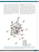
To assess the relationship among the proteins identified in the RPPA analysis, protein association network analysis was performed using STRING 10.533 on proteins identi- fied as significantly different in the AML-MSC and NL- MSC (Figure 1B). Blalock et al. used a previous version of String software to map the nuclear interactome.36 In cases where a family of proteins was identified (e.g. PPP2R2 set and HSP90 set), a representative member was included in the analysis. With the exception of PDK-1 all proteins are interconnected at least through one association (Figure 3). This finding suggests that there is an interconnection between the various proteins that are distinctly expressed between the NL-MSC and AML-MSC groups.
Activation Inhibition
Binding
Phenotype
Catalysis
Post-translational regulation Reaction
Transcriptional regulation
Figure 3. Proteins differentially expressed in acute myeloid leukemia (AML) and normal mesenchymal stromal cell (MSC) are highly interactive. (A) String analysis was performed by String 10.0 using interactions based on “action”; available from: http://string-db.org. (B) Model of involvement of Group 1 and 2 proteins in AKT signaling. Red: proteins are members of Group 1 or 2. Yellow; proteins are non-members but possible links.
haematologica | 2018; 103(5)
817
