
Page 86 - Haematologica-5
P. 86
S.M. Kornblau et al.
Pathway analysis suggests NL-MSC have prominent adipogenic signaling while all AML-MSC populations have prominent PI3K/AKT signaling
Pathway analysis was performed using IPA. Two sepa- rate data sets were created: one with proteins from PC3 (elevated in normal MSC) and one with proteins from PC1 and 2 (elevated in AML-MSC). Proteins in group 3 are associated with adipogenesis (ninth top canonical path- way; P=2.19E-05) (Online Supplementy Figure S6). PC3 pro- teins are elevated in MSC that were presumably normal but reduced in AML-MSC suggesting differences in differ- entiation potential of MSC between NL-MSC and AML- MSC. Of the 7 proteins identified, SIRT1, FOXO1, and SMAD1 each can activate PPARβ which is a critical regu- lator of adipocyte differentiation.37-39 PC3 also displayed the strongest association with AMPK signaling (P=6.84E- 07). The top upstream regulator identified in the set of PC3 proteins was PDGFB (P=1.01E-06). PI3K/AKT path- way was highly associated with PC1 and 2 proteins, which are elevated in AML-MSC (top canonical path- ways; P=7.16E-17) (Online Supplementary Figure S7). AKT
was identified as one of the top three upstream regulators (P=1.22E-14), suggesting that signaling mediated by this survival kinase is prominent in AML-MSC.
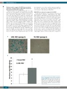
AML-MSC are senescent compared to NL-MSC
The p21 protein appears to be critical for senescence in myelodysplastic syndrome (MDS)-MSC22 and AML- MSC similar to MDS-MSC have elevated p21 (Figure 1B). This finding suggests that AML-MSC might also be more senescent than NL-MSC, as was the case in MDS-MSC.25 Senescence was observed in normal donor MSC- and AML-derived MSC using β-galactosidase staining. AML- MSC were more senescent than MSC derived from healthy donors in this representative example (Figure 4A). To account for age effects on senescence, MSC were taken from donors under 58 y. Average age of the AML patients (n=4) was 52 y and the average age of normal donors (n=5) was 47 y. Also, MSC of similar cell passage (passage 2 or 3) were used, so effects of cell passage were unlikely. As shown in Figure 4B, β-galactosidase activity was signifi- cantly higher (almost 2-fold) in AML-MSC compared to
A
B
Figure 4. Acute myeloid leukemia (AML) mesenchymal stromal cell (AML-MSC) are more senescent than nor- mal MSC (NL-MSC). (A) Representative microscopy of an AML-MSC and a normal MSC with two different slide areas. (B). Level of β-galactosidase was assessed by enzymatic assay in normal MSC (n=5) and AML-MSC (n=4). Statistical significance determined by Student t-test; *P=0.027.
818
haematologica | 2018; 103(5)
