
Page 104 - Haematologica-5
P. 104
N.A. Evensen et al.
firmed day of injection; Figure 6C) and, following confir- mation of leukemic burden on day 6, the mice were treat- ed with PBS (control) or purixan (an oral suspension form of 6-MP). Following the 10-day course of purixan treat- ment, the leukemic burden was significantly diminished in the mice harboring NT cells compared to that observed in the NT PBS treated mice (P=0.0001), suggesting that these cells were unable to survive and expand under the selective pressure of the purixan (Figure 6A and B). In con- trast, the MSH6 shRNA1 mice treated with purixan were not significantly different from the NT PBS group (P=0.828). Although purixan also had a statistically signif- icant impact on MSH6 shRNA1 cells compared to MSH6 shRNA1 PBS control (P=0.0005), these cells were able to continue proliferating under the selective pressure, unlike the NT cells (Figure 6A and B). Finally, a comparison between PBS MSH6 shRNA1 and PBS control NT cells at day 17 showed that MSH6 depleted cells also had a growth advantage in vivo (P<0.0001).
Discussion
In recent years, there has been an abundance of evi- dence demonstrating the outgrowth of clones at relapse in ALL that are associated with unique or enriched relapse
specific mutations that confer drug resistance. Some of the most common relapse specific mutations found thus far occur in NT5C2 and PRPS1 and lead to the outgrowth of thiopurine resistant clones.2,4 Our data presented here demonstrate that reduction of MSH6 in ALL also leads to decreased sensitivity to purine analogs due to a failure to initiate the apoptotic cascade directly in response to nucleotide mismatches. Even with only 50-60% reduced expression, which potentially mimics levels in patients with heterozygous loss, we demonstrate a significant decrease in sensitivity to thiopurines. Our data are consis- tent with the recent work of Diouf et al. who showed that lower levels of MSH2 in cell lines were associated with resistance to 6-TG and 6-MP. They found 11% of ALL samples showed decreased protein levels of MSH2 through copy number loss of genes controlling MSH2 degradation.17 Thus defects in MMR, including heterozy- gous deletion of MSH6, can be added to the list of genetic alterations that result in the development of resistance to purine analogs, the foundation of maintenance therapy. The variety of mutations that lead to selective outgrowth of such clones in a substantial number of patients under- scores the selective pressure of thiopurines on tumor cells.
The outgrowth of MSH6 deleted/mutated clones not found at diagnosis has been observed at relapse in malig- nant gliomas following treatment with temozolo-
AB
C
836
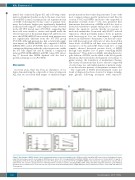
Figure 5. Knockdown of MSH6 did not lead to a mutator phenotype or increased mutation rate. (A) Microsatellite instability (MSI) was measured in diagnosis/relapse pairs that had relapse specific, heterozygous MSH6 deletions and in 697 non-targeting (NT) and MSH6 shRNA1 cells left untreated or treated with 0.05 μg/mL of 6-thioguanine (6-TG) for 5 days. (B) Mutation rate in the PIC-A gene was measured in 697 NT and MSH6 shRNA1 clones that were expanded for 2-3 weeks with or without 6-TG. The cells were analyzed for loss of GPI-dependent cell surface markers, including FLAER, CD48, CD52, and CD59 using flow cytometry. (Left) Individual mutation rates/cell divisions for each clone; the line represents mean+Standard Deviation. (Right) Mutation rates/cell divisions for three clones with and without 6- TG treatment.
haematologica | 2018; 103(5)
