
Page 95 - Haematologica-April 2018
P. 95
iAMP21 xenografts
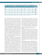
Table 2. Summary of histological and ultrastructural (EM) data for bone marrow and CNS of mice transplanted with iAMP21-ALL cells.
Xenograft
4aSLIEW
4bSLIEW 3aSLIEW 3bSLIEW 1aSLIEW 1bSLIEW 2aSLIEW 2bSLIEW
Tibia/sternum
H&E CD19 CD45 Ki67
A +ve +ve +ve
A +ve +ve +ve
A +ve -ve +ve A/B +/-ve -ve +/-ve A +ve +ve +ve A +ve +ve +ve A +ve -ve +ve
B -ve -ve -ve
Calvaria
CNS
Peak head
luminescence*
8.3x10⁷
3.4x10⁷ 3.4x10⁸ 2.3x10⁸ 3.7x10⁷ 6.4x10⁶ 3.7x10⁸ 6.3x10⁸
EM H&E
VL N/A
VL A AP A AP A VL A VL A AP A AP A
CD19 (grade)
+ve N/A
+ve 5 +ve 1-2 +ve 1-2 +ve 3-4+CP +ve 4 +ve 1
+ve 2
CNS: central nervous system; H&E: hematoxylin and eosin; EM: electron microscopy. *Units of luminescence are total flux (photons / second).
Exceptionally in xenografts of patient 1, a sharp transition in genomic architecture, involving clonal gain of CNA of three chromosomes, occurred. Chromosome 21 profiles usually remained unchanged across samples (Online Supplementary Figure S11), but interestingly in cells from patient 1, we observed structural evolution of the iAMP21 chromosome, involving a small region of copy number gain and nine distinct regions of loss of one or two copies (Figure 3A and Online Supplementary Table S8). Importantly the additional deletions did not affect two regions predict- ed to contain critical oncogenes11 but did re-define the proximal boundary of the region of highest level of ampli- fication9 from 21:32,813,553-37 to 21:33,949,423. By FISH, we confirmed that the RUNX1 and APP gene regions were maintained at the same level of amplification and reduced in copy number from three to one, respectively. Additional rearrangements included bi-allelic deletion of the short arm of chromosome 9 (9p), resulting in homozygous loss of CDKN2A/B, as confirmed by MLPA (Online Supplementary Table S9), and mono-allelic deletion of 3p, involving the CMTM genes 6-8. SNP6.0 array and FISH provided no evi- dence of these CNA prior to their emergence in 2°1a. However, as previously reported, two reads in whole genome sequencing data were consistent with the pres- ence of a minor clone carrying the chromosome 3 deletion in the patient’s cells at presentation.11
Suggestive of convergent clonal evolution and highlight- ing the relevance of specific chromosomal regions to dis- ease progression, several were targeted by different abnor- malities segregated in xenografts from the same patient. Consistent with an oncogenic role for genes on Xp, patient 2 carried competing sub-clones marked by gain of a whole X chromosome or isochromosome Xp (Figure 3B). Whether emergence of a focal deletion of Xp, involving the zinc finger genes, ZNF157 and ZNF41, was related to the presence of the larger scale CNA remains unclear but they were unlikely to have been driven by CRLF2 overexpres- sion, as genetic analysis ruled out rearrangement of this locus in the patient’s sample.25 In patient 3 large overlap- ping deletions of 12p, both involving ETV6, were segregat- ed (Figure 3C). Although no patient’s material was avail- able, differences in CNA involving the long arm of chro- mosome 10 were identified in xenografts from a relapse sample of patient 4 (Figure 3D). Strongly indicative of con- vergent evolution and hence a role in leukemia progres- sion, the same focal bi-allelic deletion, involving PIK3AP1 (BCAP) and LCOR (C10orf21), was nested within two dis-
tinctly different, large, mono-allelic 10q deletions. Evidently of independent origin, as it was detected only in a single xenograft, one of the large deletions also harbored a second likely co-operating focal bi-allelic deletion that resulted in loss of BLNK. Lastly, passage of patient 5 cells in a primary xenograft resulted in concomitant loss of copy number neutral loss of heterozygosity of 12q, with pro- gression from sub-clonal to clonal deletion of a region of 12q containing SH2B3 (Figure 3E). Comparison between relapse and the xenograft showed no overlap in progres- sion of specific CNA, although interestingly the EBF1 gene was targeted by different deletions in the two samples.
Mutations affecting the RAS pathway drive clonal expansion
To investigate progression of mutations activating the RAS pathway previously identified in patient 113 we per- formed whole exome sequencing of selected and Sanger sequencing of all derived xenografts (Figure 4A). Interestingly, while an NF1 mutation remained clonal in all samples, two different mutations affecting NRAS and one of KRAS marked a dramatic clonal evolution. Remarkably the KRAS mutation, present as a dominant clone in both primary and one secondary xenograft was undetectable at a read depth of over 6000 in the presentation sample and also undetected in other xenografts which instead carried a dominant NRAS mutation detected in only 1% of reads at presentation. NRAS and FLIT3 mutations detected at pres- entation in patients 3 and 5, respectively, were clonal in xenografts while the FLIT3 mutation was lost at relapse (Figure 4B,C).
Transcriptional environment associated with deletions
Anticipating that xenograft preparations, in contrast to patients’ samples, would be free of non-leukemic human cells, we used them to investigate the transcriptional envi- ronment associated with bi-allelic deletions (Table 3). By comparison with non-deleted samples, and confirming clonal dominance of the chromosome 10 deletions, we saw marked reductions in the levels of transcription of PI3KAP1, LCOR and BLNK. Loss of the BLNK genomic region also resulted in silencing of DNTT and OPALIN. Three of the four sequenced xenografts carried bi-allelic 9p deletions affecting CDKN1A/B. Indicating strong pressure for clonal selection of bi-allelic loss, read counts within this region were reduced to zero in deleted cases. Interestingly two focal bi-allelic deletions were associated with silencing
haematologica | 2018; 103(4)
639
