
Page 49 - Haematologica-April 2018
P. 49
Hepcidin and hyperferritinemia in Gaucher disease
regarding iron parameters was compared between them (Figure 7A and B). In Patient 1, an 18-year-old woman, the ferritin level decreased dramatically after only five months of ERT, and this decline was maintained continuously over time (Figure 7A). Consequently, the TS began to increase, reaching a maximal level (approximately 40%) at 10 months post ERT, and then returned to a steady state value of approximately 30%. The kinetics of systemic hepcidin secretion was again positive and transient with a maximal level at 18 months post ERT, allowing the TS to reach normal values. This controlled iron release was ben- eficial for restoring hemoglobin synthesis because its serum level requires ten months post ERT to stabilize. Similar kinetic patterns were confirmed in another young treated patient (Online Supplementary Figure S4), although the TS values and intermediate time data were missing. Patient 2 was a woman who received ERT for the first time at 52 years of age. Interestingly, the decrease in the ferritin level was not evident as late as 15 months post ERT. The kinetic patterns of the other parameters were similar to those of Patient 1 (Figure 7B).
Finally, we analyzed the correlation between the hep- cidin and ferritin values in untreated and treated GD1 patients. Compared with that in untreated patients, the correlation between both parameters was significantly improved under ERT with a higher slope (Figure 7C). Indeed, the hepcidin response levels were more adapted toward the ferritin values, confirming the restored sys- temic hepcidin control on iron metabolism. Furthermore, this recovery of controlled iron metabolism was associat- ed with a reduction in the soluble transferrin receptor level in treated patients (Figure 6E).
Discussion
This study demonstrated for the first time that hyperfer- ritinemia observed in GD1 patients is related to abnormal iron metabolism and the sequestration of iron in Gaucher cells. ERT allows for the release of iron, improves the iron status, and corrects the anemia. In addition to spleen dys- function, bone marrow infiltration by Gaucher cells and ineffective erythropoiesis, part of the anemia can likely be explained by this abnormal iron metabolism.
From a large cohort of 90 GD1 patients, we showed that most of them (65%) exhibited high levels of serum fer- ritin, with one-third presenting moderate anemia. These results are in line with previous data.9-11 Serum iron and TS were in a normal range as previously shown in adult GD1 patients.4,9 However, the pediatric population exhibited decreased iron release into plasma as shown by a low TS and significant anemia. Indeed, children were the most affected patients, most likely because the iron require- ments are very high during childhood due to the high energy intake and active erythropoiesis, limiting the body pool of iron.31 In our cohort of GD patients, hyperferritine- mia was not related to an obvious systemic inflammation because neither CRP nor IL-6 were detected. Furthermore, there was no difference in hepcidin level, which is also induced by inflammation,32,33 between the groups of GD patients with and without hyperferritinemia. A normal serum hepcidin concentration despite hyperferritinemia has already been reported by Lorenz et al. in a limited number of GD1 patients (n=11).11 Although Medrano- Engay et al. reported increased hepcidin levels in 3 untreat- ed GD1 patients,10 our data indicate that the ferritin levels
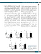
ABC
DE
Figure 5. Impact of CBE treatment on J774 inflammatory profile. IL-1β, IL-10, MCP-1, CCL-5 and TNF-α were quantified in super- natant of J774 cells after incubation with (+CBE) and without (-CBE) CBE for 96 hours (h). Mann-Whitney test was used to compare cytokine levels.
haematologica | 2018; 103(4)
593
