
Page 47 - Haematologica-April 2018
P. 47
Hepcidin and hyperferritinemia in Gaucher disease
lational downregulation of FPN may involve hepcidin and macrophages can produce hepcidin locally, we quantified the expression level of the Hamp gene encoding hepcidin in J774 cells under the same conditions. We found that inhibition of GCerase activity significantly increased the mRNA level of hepcidin, which may lead, at least in part, to a reduction in FPN protein via an autocrine-paracrine interaction and resulting in iron retention in these cells (Figure 4C). Since hepcidin in liver was found to be induced by inflammatory cytokines, likely by IL-6 or IL- 1β, we also explored the J774 cells inflammatory profile by quantifying secreted cytokines and chemokines in their supernatant. under CBE treatment, the levels of IL-1β, MCP-1 and CCL-5 levels were increased, the levels of IL-
AB
10 and TNF-α levels were not affected, and INF-g, IL-6 and IL-12 remained under limit of detection (Figure 5). Thus, one can postulate that increased hepcidin in CBE-treated J774 macrophages is dependent on a local inflammatory environment.
The improvement of biological abnormalities by enzyme replacement therapy confirms the local iron sequestration hypothesis
Serum ferritin was significantly reduced in children (104 vs. 287 mg/L; P=0.008). A trend toward reduced values was observed in adults (Table 1). Hyperferritinemia was observed in 42% of treated rather than 65% of untreated patients (P=0.02) (Online Supplementary Table S2). This
Figure 2. Serum hepcidin level and transferrin saturation (TS) are inde- pendent of the ferritin status. Serum hepcidin (A) and TS (B) levels were measured in the normal fer- ritin (NF) and hyperferritin (HF) untreated patient groups. The means are represented by the hori- zontal line. According to a two-tailed Mann-Whitney test, no significant differences were observed.
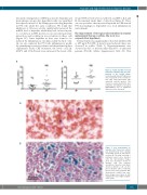
A
B
Figure 3. Iron sequestration in Gaucher cells. Tissue iron content was determined by Perl’s staining of medullar smear (A) and spleen sections (B). The large cells with a laminated aspect were Gaucher cells. The representative images show iron deposition mostly in Gaucher cells. The classical description of Gaucher cells (black arrows) is limited to cells 20-100 μm in diameter with eccentrically placed nuclei and cytoplasm with characteristic crinkles and stria- tions. Images were taken at 20X magnification and a higher magnifi- cation (40X).
haematologica | 2018; 103(4)
591
