
Page 51 - Haematologica-April 2018
P. 51
Hepcidin and hyperferritinemia in Gaucher disease
ABC
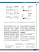
Figure 7. Recovery of hepcidin control on iron metabolism in treated Gaucher disease (GD) patients. (A and B) Time course of iron-related parameters in 2 treated patients from enzyme replacement therapy (ERT) initiation until 18-25 months after ERT initiation. Patient 1: 18-year-old woman; Patient 2: 52-year-old woman. (C) According to the Spearman test, hepcidin/ferritin correlation in the untreated and treated patients r and P were shown for each condition and slope of linear regres- sion.
levels under ERT may also be partly due to this iron avail- ability. This was confirmed by the lower level of soluble transferrin receptor in treated patients. Similarly, an increased number of reticulocytes in treated patients would have helped to highlight this mechanism, but unfortunately these data were not available. However, iron restriction may not be the only cause of GD anemia, as we have already shown that GD patients exhibit an ineffective erythropoiesis,7 as well as altered morphologi- cal properties of RBCs that could accelerate their ery- throphagocytosis.43,44 In this study, the efficacy of ERT was particularly demonstrated in the pediatric population in which the iron requirement is high. Likewise, the low fer- ritin responsiveness in Patient 2 under ERT may imply a low efficacy of this treatment in older patients, but this hypothesis requires additional studies on a larger number of patients. Interestingly, the level of serum hepcidin was increased after ERT. This is probably to fight against iron excess due to the improvement of iron release from Gaucher cells, and to regain a balanced iron status.
In conclusion, our study revealed for the first time that hyperferritinemia in GD is related to the sequestration of iron in Gaucher cells due to the downregulation of the
iron exporter ferroportin. Hepcidin from macrophages, rather than the systemic peptide, seems to be responsible for this FPN repression. ERT restored the iron availability and improved the anemia in these patients. Overall, these results provide new perspectives for better management of GD patients. Indeed, the measurement of liver iron con- tent by magnetic resonance imaging and the analysis of iron status must be systematically evaluated to prevent iron deficiency in patients.
Acknowledgments
We are very grateful to the patients who kindly contributed to this study. Inserm and Paris Diderot University, the Laboratory of excellence, GR-Ex, Paris, France, supported this work.
Funding
The labex GR-Ex, reference ANR-11-LABX-0051 is funded by the program “Investissements d’Avenir” of the French National Research Agency, reference ANR-11-IDEX-0005-02. We thank Nathalie Dessendier, Nicolas Ducrot and Olivier Thibaudeau for their technical support, Samira Benadda for the confocal microscope support, and Karima Yousfi for providing assistance in collecting the patient data.
References
1. Brady RO, Kanfer JN, Shapiro D. Metabolism of glucocerebrosides. II. Evidence of an enzymatic deficiency in Gaucher’s disease. Biochem Biophys Res Commun. 1965;18:221-225.
2. Grabowski GA. Phenotype, diagnosis, and treatment of Gaucher’s disease. Lancet Lond Engl. 2008;372(9645):1263-1271.
3. Charrow J, Andersson HC, Kaplan P, et al. The Gaucher registry: demographics and disease characteristics of 1698 patients with Gaucher disease. Arch Intern Med. 2000;160(18):2835-2843.
4. Stein P, Yu H, Jain D, Mistry PK. Hyperferritinemia and iron overload in type 1 Gaucher disease. Am J Hematol. 2010;85(7):472–476.
5. Regenboog M, van Kuilenburg ABP, Verheij J, Swinkels DW, Hollak CEM. Hyperferritinemia and iron metabolism in Gaucher disease: Potential pathophysio-
haematologica | 2018; 103(4)
595
