
Page 168 - Haematologica-April 2018
P. 168
D. Belloni et al.
mediated drug resistance (SFM-DR) and cell adhesion- mediated drug resistance (CAM-DR).27,28 To assess the impact of 3D microenvironment on bortezomib sensitivi- ty, we performed parallel experiments in 2D versus 3D using HS-5 cells as stroma. Co-culture of MM1.S with HS- 5 cells in 2D conditions conferred higher resistance to bortezomib, compared to MM.1S alone (Figure 4A, left). Significantly, resistance to bortezomib was even more evi- dent when MM1.S were cultured with HS-5 cells in biore- actor, underscoring the role of 3D architecture per se in determining the impact of drugs (Figure 4A, left). The pro- tective effect exerted by HS-5 cells was negligible with the RPMI.8226 cell line (Figure 4A, middle), in agreement with their reduced adhesion to stroma (Figure 2B). Protection conferred by HS-5 cells was not restricted to bortezomib- treated MM1.S since it was also observed with other cell lines (bortezomib-treated U266) (Figure 4A, right) and other drugs (melphalan) (Figure 4B), particularly in 3D conditions.
HS-5 cells in principle can provide both mechanisms of drug resistance, since they release consistent amounts of
IL-6 (Figure 3D) and serve as support to MM cells. Among tumor-stroma cell-cell interactions, we addressed as a par- adigm the VLA-4/VCAM1 molecular pathway, which is considered to play a central role.27,28 In experiments per- formed in 2D conditions, primary VCAM1+ BMSC (Online Supplementary Figure S3A) from MM patients promoted adhesion at least in part via the counter ligand VLA-4 (Online Supplementary Figure S3B), as demonstrated by inhibition experiments with the specific α4 blocking anti- body natalizumab,29 and accordingly conferred to MM1.S cells higher resistance to bortezomib compared to the VCAM1 negative HS-5 cell line (Online Supplementary Figure S3C); the release in culture supernatants of β2-microglobulin and of lactate dehydrogenase (LDH), the latter bona fide expression of bortezomib cytotoxicity, par- alleled this response (Online Supplementary Figure S3C).
We then modeled the VLA-4/VCAM1 pair using the murine fibroblast L-VCAM transfectant (Figure 4C). The capability of VCAM1 expressed by the transfectants (Online Supplementary Figure S3A) to engage specific inter- actions was demonstrated by inhibition experiments
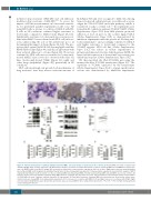
A
BCD
EF
712
Figure 5. Survival and functions of primary multiple myeloma (MM) cells and stroma are promoted in the scaffold. (A) Primary MM cells inside scaffold retain CD138 and light chain (κ chain) expression. (B) Western blot analyses performed on parallel 2D and 3D co-cultures of primary MM cells and MM bone marrow stro- mal cells (BMSC); pAkt, total Akt, β1 integrin (β1) and actin are depicted in a representative experiment (left) out of three. Right panels represent mean±Standard Error of Mean (SEM) of β1 integrin/actin and pAkt/total Akt ratios in three independent experiments. (C) Representative experiment of western blot analysis of STAT3 and Akt phosphorylation in 2D versus 3D co-cultures of primary MM cells and primary BMSC. Freshly isolated primary MM cells and BMSC are used as controls. (D) Input (t0) and recovered (t7) number of primary CD38+ MM cells from 7 patients after 3D culture for seven days. Dotted lines represent input and recovered number of CD38+ MM cells from 3 patients cultured in parallel 2D experiments. (E) Specialized functions of primary MM cells and stroma are measured in culture super- natants. Data are mean±SEM of six independent experiments. (F) IL-6 release in co-cultures with BMSC (left) and Ang-2 release in co-cultures with HUVEC (right) were determined by ELISA in parallel 2D and 3D experiments. Data are mean±SEM of three independent experiments. *P≤ 0.05; **P≤0.01. H&E: hematoxylin and eosin staining. Bar=100 mm.
haematologica | 2018; 103(4)
