
Page 167 - Haematologica-April 2018
P. 167
3D reconstructed myeloma microenvironments
(nude scaffold) of stroma; this was particularly evident with MM1.S cells (Figure 2C). Accordingly, immunohis- tochemistry (IHC) indicated that both MM1.S and RPMI.8266 cells entered, were homogeneously distributed and proliferated inside the scaffolds, prevalently when pre-seeded with the HS-5 stromal cell line (Figure 2D). Other cell types within the MM BM microenvironment, including endothelial cells and osteoblasts, are increasing- ly recognized as participating in MM pathogenesis and progression.12,24 We then exploited our system to model MM cells-HUVEC and MM cells-osteoblasts co-cultures. The latter were obtained through bone differentiation of BMSC, as reported.18 Upon culture with osteogenic differ- entiation medium, BMSC underwent morphological changes, increased mineralization and acquired Alizarin staining (Online Supplementary Figure S2A-C). Moreover, parallel cultures performed with differentiated BMSC in 2D and, particularly in 3D conditions, showed alkaline phosphatase release (Online Supplementary Figure S2D). Notably, scaffolds seeded with bone differentiated BMSC and with CD31+HUVEC also supported MM cells homing and permanence (Figure 2D, right panels).
Development of intimate cell-cell contacts between MM cells and microenvironment was visualized at scan- ning electron microscopy analysis (Figure 2E, middle panel) showing that MM cells acquired a flatter morphol- ogy over the stroma, consistent with the induction of
adhesion-mediated cytoskeletal rearrangement. Conversely, MM cells exhibited a round shape with few contact points over nude scaffolds (Figure 2E, left panel). Interestingly, the entire 3D cell surface of some MM cells was embedded when HUVEC were used as stroma, sug- gesting that cell-cell interactions may dynamically evolve upon contacts (Figure 2E, right panel). The recapitulated physical interactions resulted in the activation of pro-sur- vival signals, as indicated by up-regulated Akt phosphory- lation in MM cell line-stromal cell co-cultures in 3D com- pared to 2D co-cultures (Figure 3A and B); instead, pAkt expression was negligible in isolated HS-5 cells. Similarly, the expression of survivin was also increased in 3D co-cul- tures (Figure 3A).25,26 Specialized functions of both MM cells and stroma were detectable in culture supernatants, including β2-microglobulin (Figure 3C) and growth factor release. The latter varied according to the stromal ele- ments coating the scaffolds: Ang-2 was related to the pres- ence of HUVEC,20 while IL-6, VEGF and FGF were detectable in all co-cultures (Figure 3D).
Response to anti-myeloma drugs of MM cell lines inside the surrogate microenvironment
Tumor-stroma interactions affect MM cell behavior, including survival and drug resistance. The latter is induced via two overlapping pathways, i.e. soluble factor-
AB
CDE
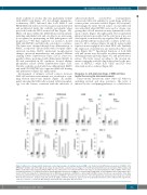
Figure 4. Adhesion to stroma and 3D architecture confer drug resistance to multiple myeloma (MM) cells. (A) Bortezomib (BTZ)-induced apoptosis in MM1.S (left), RPMI.8226 (middle) and U266 (right) in 2D versus 3D experiments, evaluated as percentage (%) of CD38+annexinV (AnnV)+ by flow cytometric (FACS) analysis. (B) MM1.S death induced by melphalan (Melph). MM cells were either alone or co-cultured with HS-5 cells. Data are mean±Standard Error of Mean (SEM) of three inde- pendent experiments. (C) Comparison between 2D/3D BTZ-induced apoptosis of MM1.S co-cultured with L-VCAM. Data are mean±SEM of three independent exper- iments. (D) The blocking effect of natalizumab (nat) on BTZ-induced apoptosis on MM1.S co-cultured with L-VCAM is shown. Results are representative of two inde- pendent experiments. (E) Dexamethasone-induced apoptosis in MM1.S cells in the presence/absence of IL-6 (10 ng/mL) was evaluated as percentage (%) of CD38+ AnnV+ by flow cytometric analysis in 2D (left) and in 3D (right) conditions. Results are expressed as mean±Standard Deviation of three independent experiments. *P≤0.05; **P≤0.01; ***P≤0.001. WT: wild-type non transfected counterpart; NT: not treated.
haematologica | 2018; 103(4)
711
