
Page 165 - Haematologica-April 2018
P. 165
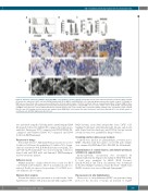
3D reconstructed myeloma microenvironments
ABC
D
E
Figure 2. Stroma is required for multiple myeloma (MM) cell permanence inside scaffolds. β1-integrin and α4 chain expression by flow cytometric (FACS) analysis (A) and in vitro adhesion to HS-5 cells and VCAM1 transfectant (B) of MM1.S and RPMI.8226 cells. Gray histograms represent the isotype controls. (C) Number of MM cells recovered from nude or pre-seeded scaffolds after 24 hours (input number =500x103/scaffold). Data are mean±Standard Error of Mean (SEM) of three independent experiments. (D) Immunohistochemistry (IHC) showing proliferating (Ki67+) CD138+ MM cells over a layer of HS-5 cells or CD31+HUVEC. CD138 staining of MM1.S in the presence of bone-differentiated bone marrow stromal cells is also shown. Insert represents alizarin staining of the osteoblasts-coated scaffold. Bar=100 mm. (E) Scanning electron microscopy analysis shows RPMI.8226 cells without (left panel and insert, bar=2 mm) and with HS-5 cells (middle panel) or endothelial cells (HUVEC) (right panel). Bar=20 mm.
was performed using the following mAbs: anti-β1integrin/CD29 (BioLegend), followed by ALEXA-488-conjugated goat anti-mouse antibodies (Invitrogen); FITC-conjugated anti-VLA4/CD49d; PE- conjugated anti-VCAM-1/CD106, PC7-conjugated anti-CD38 (both from BD-Pharmingen).
Response to drugs
Bortezomib (Velcade®, Millenium Pharmaceuticals) was used at 10 nM for 24-48 hours (h), melphalan at 1.2 nM for 72 h, dexam- ethasone at 20 nM for 48 h; IL-6 (R&D Systems) at 10 ng/mL. The anti-VLA-4 mAb natalizumab19 was used at 10 mg/mL. Cells were stained with anti-CD38 mAb and Annexin-V (BD-Pharmingen) for flow cytometric analysis.
Adhesion assay
Multiple myeloma cell lines (200x103) were seeded over HS-5 or L-VCAM1 in 24-well plates. After 3 h, non-adherent cells were removed. Results are expressed as percentage (%) of CD38+ recov- ered adherent cells over input.
Western blot analysis
Western blot analysis was performed as described in the Online Supplementary Methods with polyclonal anti-pAkt (against S473,
R&D Systems), monoclonal anti-pan-Akt (clone C67E7, Cell- Signaling Technology), anti-β1integrin mAb (abcam), anti-β-actin mAb (Santa Cruz Biotechnology), anti-STAT3/p-Stat and survivin (abcam). Proteins were quantified by ImageJ software.20
Scanning electron microscopy analysis
Scaffolds were fixed in 2% glutaraldehyde, post-fixed in 1% OsO4, dehydrated and then sputter coated with gold. Samples were examined by FEI/Philips XL-30 SEM (FEI, the Netherlands).
Determination of soluble factors and metallo-proteasic activities in supernatants
β2-microglobulin concentration was determined by immunonephelometry. Angiopoietin-2 (Ang-2), VEGF, FGF and IL- 6 levels were quantified by ELISA (R&D Systems). IL-1β,IL-8/CXCL-8 and TGF-β concentrations were determined by Bio-Plex Multiple-Cytokines Assays (Bio-Rad).21 MMP-2 and MMP-9 activities were assessed through Zymography.16
Fluorescence in situ hybridization
Fluorescence in situ hybridization (FISH)22 was performed using
probes for the detection of trisomy 12, deletions of 11q22.3
haematologica | 2018; 103(4)
709
