
Page 118 - Haematologica-April 2018
P. 118
Z. Li et al.
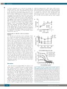
of 3C12C (10 mg/ml) on ice followed by washing off unbound antibody. Cells were then cultured without 3C12C for up to two hours. The remaining 3C12C bound on the cell surface were detected by a secondary anti- human antibody. Though L428 and HDLM2 have a lower level of surface CD83 expression compared to KM-H2, our analysis showed that the 3C12C level on the surface of HDLM2 reduced much faster than on L428, while the 3C12C bound to L428 were far more stable (Figure 6B). This suggested 3C12C was rapidly internalized in KM-H2 and HDLM2, while 3C12C was internalized slower in L428. To investigate further potential therapeutic applica- tions, we generated a 3C12C toxin-conjugate (3C12C- MMAE). In vitro, 3C12C-MMAE killed CD83+ KM-H2 cells most efficiently, followed by HDLM2 and L428, while CD83– HL-60 cells were the least sensitive to 3C12C-MMAE (Figure 6C and Online Supplementary Figure S3). In addition, the intracellular CD83 level in HDLM2 was much higher than L428, lending to more sensitivity of the HDLM2 to the killing of 3C12C-MMAE (Online Supplementary Figure S1).
Administration of 3C12C is safe in non-human primates
To “de-risk” the antibodies before advancing 3C12C into a clinical trial, we performed pre-clinical dose-escala- tion studies of 3C12C in non-human primates. Five baboons were injected intravenously with 3C12C (1, 5, 10 mg/kg on d0, 7, 14, and 21). No adverse clinical events were recorded during follow up for 84 days. We assessed blood counts and biochemistry weekly, and monitored different immune cell populations by flow cytometry or immune histology. Administration of 3C12C did not affect blood cell counts (white blood cells [WBC], red blood cells [RBC], and platelets), liver (ALP and AST) or kidney (creatinine) function (Online Supplementary Figure S4). The total T-cell number, and ratio of CD4+/CD8+ T cells all remained normal up to day 84 (data not shown). However, there was evidence of 3C12C efficacy in that CD1c+DC counts were reduced. We found that baboon blood B cells expressed CD83 as human B cells (data not shown), and reductions in blood B cells were noted by flow cytometry (Figure 7A). In addition, B-cell areas in lymph nodes were reduced in the 3C12C-treated animals (10mg/kg) compared to the control animals (human IgG 10mg/kg) (Figure 7B).
Discussion
HL is driven by the malignant HRS cell, which are of B lineage origin.31,32 A significant number of patients experi- ence relapsed/refractory disease following first-line chemotherapy.33 Less toxic treatments for relapsed/refrac- tory HL would be highly desirable, as exemplified by the introduction of the anti-CD30 antibody drug conjugate (brentuximab).10,34 In the study herein, we were able to identify sCD83 as a new potential biomarker for HL, and CD83 as a target for a therapeutic mAb and derivatives.
CD83 was first described on activated B cells and we
19 originally detected CD83 on HL using frozen sections.
Our ability to stain paraffin embedded lymph node biopsy samples of HL patients encouraged this study and allows for the assessment of CD83 expression in routine clinical practice. CD83 was highly expressed on HRS cells with a
different staining pattern to CD30. Thus, CD83 is poten- tially another diagnostic marker of HL. More importantly, this work suggests that the majority of HL patients might be suitable for a therapeutic mAb targeting CD83. An anti-CD83 mAb may also work synergistically with chemotherapy, which is similar to the treatment of stage III or IV HL with anti-CD30 antibody drug conjugate
Figure 6. 3C12C and 3C12C conjugation with monomethyl auristatin E (3C12C-MMAE) kill Hodgkin lymphoma (HL) cell lines in vitro. (A) Target cells KM-H2, L428 or HDLM2, labeled with Calcein-AM were co-cultured with effector cells (human PBMC) at effector: target ratio of 25:1 with increasing 3C12C con- centration from 0 mg/ml to 1 mg/ml at 37°C for three hours. Supernatant was collected for fluorescence reading (excitation 485nm, emission 538nm) of released Calcein. Antibody (Ab)-dependent cell cytotoxicity was calculated (n=3). (B) HL cells were cultured in 3C12C saturation concentration (10 mg/ml) on ice followed by intensive washing and culture without 3C12C from 0-2 hours. The remaining levels of 3C12C bound on the cell surface were detected by a secondary anti-human antibody with flow cytometry. The remaining surface level of 3C12C on KM-H2, L428 and HDLM2 was normalized to the level of time 0. (n=3). (C) CD83+ KM-H2, L428, HDLM2 or CD83–HL-60 cells were cultured with different concentrations of 3C12C-MMAE for three days before determining viable cells by 7-amino-actinomycin D (7AAD) staining with flow cytometry. The half maximal inhibitory concentration (IC50) is shown. Data was from one of four representative experiments.
A
B
C
662
haematologica | 2018; 103(4)
