
Page 114 - Haematologica-April 2018
P. 114
Z. Li et al.
middle. Interestingly, strong CD83 expression on HRS cells was found in two out of three relapsed HL (Online Supplementary Table S1). Epstein-Barr virus (EBV) infection is associated with an increasing risk of developing EBV- positive HL. A number of viral products, including EBV nuclear antigens (EBNA), EBV latent membrane proteins (LMP1 and LMP2) and EBV encoding small ribonucleic acids (RNA; EBER) have been implicated. LMP1 induced CD83 in EBV-infected human B cells by activation of NF- kB.29 CD83/LMP1 has been reported to be correlated in MC HL, but not for NS HL.23 By in situ staining of EBER of 35 HL samples, we found that seven HL were EBER posi-
tive, including 2/7(28.6%) MC and 3/22 (13.6%) NS HL (Figure 2D). On six out of seven EBER positive HL sam- ples, CD83 staining of HRS were strong or moderate (Online Supplementary Table S1).
CD83 is trogocytosed from HL cells to T cells
We found previously that CD83 was able to transfer from the membrane of DC to T cells via trogocytosis.15 Similar trogocytosis was observed to occur between HL cell lines and T cells. When these two cell types were co- cultured for four hours, CD83 surface expression was detected on 5-15% of T cells (Figure 3A,B), whereas no
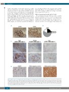
AB
C
D
658
Figure 2. CD83 is expressed on Hodgkin and Reed-Sternberg (HRS) cells in Hodgkin lymphoma patients. (A) CD83 and CD30 expression (brown) on paraffin-embed- ded lymph node biopsy samples of HL was imaged by microscopy with ×200 magnification. One representative sample of 35 biopsies shown. (B) Pie chart analysis of CD83 expression level in HRS cells of HL patients (n=35). High: CD83 positive in >90% HRS cells; middle: 10-90% CD83+in HRS cells; low: 10% CD83+ in HRS cells. One representative sample of each group is shown in (C), upper panel: original magnification ×40, lower panel shown with high amplification (×200). Arrows indicate HRS cells expressing CD83. (D) Epstein-Barr virus encoding small ribonucleic acids (RNA; EBER) in 35 HL biopsies were detected by in situ hybridization; one of the seven EBER positive samples is shown.
haematologica | 2018; 103(4)
