
Page 113 - Haematologica-April 2018
P. 113
CD83 and Hodgkin lymphoma
Results
CD83 is expressed on HL cell lines and HRS cells in lymph node biopsies of HL patients
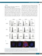
Expression of CD83 was analyzed using the mouse anti- human antibodies HB15a, HB15e and potential therapeu- tic human anti-human CD83 antibody 3C12C.25 KM-H2 cells expressed the most expressive CD38 cell surface, stained as it was with all three anti-CD83 antibodies, whilst the L428 and HDLM2 lines expressed less CD83. All three lines expressed CD30 (Figure 1A). This data was confirmed by confocal CD83 staining on KM-H2 cells (Figure 1B), detection of CD83 messenger ribonucleic acid (mRNA) transcripts by reverse transcription polymerase chain reaction (RT-PCR) and intracellular CD83 expres- sion in the three HL lines (Online Supplementary Figure S1).
Next, CD83 expression was analyzed on the paraffin- embedded lymph node biopsies of 35 HL patients (Table 1). The HRS cells were identified as CD30+ (Figure 2A). Of note, 8/35 (22.9%) biopsies expressed high levels of CD83 on the HRS cells (>90% positive), 21/35 (60%) expressed middle levels (10-90% positive), and 6/35 (17.1%) expressed low levels of CD83 (<10% positive) (Figure 2B,C). Of the 29 biopsies with high or middle expression, 21 (72.4%) had strong or moderate intensity, while 8/29 (27.6%) showed weak intensity of CD83 on HRS cells (Online Supplementary Table S1). The subtype analysis showed that 16/21 (79.2%) of HRS cells in nodular sclero- sis (NS) HL were CD83 high or middle, and 85.7% were CD83 high or middle in mixed cellularity (MC) HL. Most (20/22, 90.9%) of stage I-II HL were CD83 high or middle, and 9/13 (69.2%) HL in stage III-IV were CD83 high or
A
B
Figure 1. CD83 is expressed on Hodgkin lymphoma cell lines. (A) Expression of CD83 was analyzed by flow cytometry on KM-H2, L428 and HDLM2 cell lines, which were stained with HB15a-fluorescein isothiocyanate (FITC), HB15e-FITC or 3C12C-FITC anti-CD83 mAbs, respectively. Gray histograms represent isotype control, while open histograms represent anti-CD83 antibodies. CD30 staining was used as a positive control. These data are representative of three independent experiments with comparable results. (B) CD83 expression (red) on KM-H2 cells with HB15a, HB15e or 3C12C mAb were imaged by confocal microscopy. Nuclei were stained with 4',6-diamidino-2-phenylindole (DAPI; blue). Human IgG1 was used as control for 3C12C mAb. Scale bar: 5μm.
haematologica | 2018; 103(4)
657
