
Page 98 - 2020_08-Haematologica-web
P. 98
S. Wang et al.
Loss of Tfr1 in HSC causes impaired development of various cell lineages in the FL
Our finding that cKO pup were anemic at birth (Figure 4A) suggests that hematopoiesis is impaired in prenatal development. Moreover, definitive hematopoiesis involves the colonization of the FL, thymus, spleen, and ultimately the BM.5,6 Therefore, we harvested FL cells from cKO and control embryos at E14.5, E16.5, and E18.5 and performed hematopoietic phenotyping using multi- color flow cytometry. Our analysis revealed no difference between cKO and control embryos at E14.5. However, during later stages of development, the cKO embryos became progressively smaller and paler than control sib- lings (Figure 4B). The most prominent difference was a significant decrease in lineage-positive (Lin+) cells begin- ning at E16.5 and becoming much more pronounced by E18.5 (Figure 4C-D), indicating that some cell lineages were severely impaired.
To examine this effect in further detail, we measured the development of erythroid, myeloid, and lymphoid cells in FL. With respect to erythropoiesis, we found no effect at E14.5. In contrast, R1 and R3 increased at both E16.5 and
E18.5, and R4 and R5 significantly reduced at E18.5 (Figure 4E); thus, progressive erythropenia in cKO embryos occurs after E14.5. With respect to myelopoiesis and lymphogen- esis, we found reduced numbers of both Mac1+Gr1+ gran- ulocytes and CD3+ T cells in the cKO embryos beginning at E14.5, whereas the number of CD19+ B cells was similar between cKO and control embryos (Figure 4F). These results indicate that the loss of Tfr1 in HSC causes impaired development of myeloid cells, erythrocytes, and T cells but spares B-cell development in the FL, consistent with our results obtained in neonatal mice.
Given the inability of cKO mice to produce several mature cell lineages, we examined HSPC in the FL of cKO and control embryos at E16.5, the gestational stage in which Lin+ cells dramatically decreased. Interestingly, we found no difference between cKO and control embryos, with the exception of a slight albeit significant increase in granulocyte/macrophage progenitors (Figure 4G). These data suggest that in the FL, Tfr1 preferentially functions at the progenitor/precursor stage rather than the HSC stage, consistent with our findings shown in Figure 1 and Online Supplementary Figure S1.
ABCD
E
F
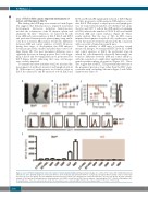
Figure 2. Loss of Tfr1 in hematopoietic stem cells results in early postnatal lethality. (A) Representative images of a control ( Tfr1fl/fl) and a Trf1 conditional knockout (cKO) mouse at P6. (B) Body weight of control and cKO mice at the indicated days after birth (n=4 for each group). (C) Kaplan–Meier survival curve for control and cKO mice (n=7 for each group). (D) Tfr1 mRNA was measured in bone marrow cells obtained from control and cKO mice (n=3 for each group). (E) Flow cytometric analysis of Tfr1 expression in hematopoietic stem/progenitor cells (HSPC) in fetal liver of E14.5 mouse embryos. Open histogram: Tfr1fl/fl control; red histogram: cKO. (F) Quantification of the flow cytometry data in (D). The mean fluorescence intensity (MFI) of Tfr1 in the indicated HSPC. **P<0.01; ***P<0.001.
P=0.0265
2074
