
Page 97 - 2020_08-Haematologica-web
P. 97
The critical function of Tfr1 in hematopoiesis
compared to control littermates (Figure 3B); in contrast, the number of B220+ cells in the liver and spleen was sim- ilar between cKO and control mice (Figure 3C). We also found significantly fewer cells in the thymus of cKO mice (Figure 3D), particularly CD4+CD8+ T cells (Figure 3E). Taken together, these results indicate that Tfr1 is required for the generation of T cells, macrophages, granulocytes, and erythrocytes, but is not required for the production of B cells or platelets.
Loss of Tfr1 impairs hematopoiesis in the BM
During late embryogenesis, HSC populate the BM, pro- viding stem cells and progenitor cells for all blood cell lin-
eages; thus, during postnatal development the BM becomes the primary site of hematopoiesis.23 Hematoxylin and eosin staining revealed significantly fewer cells in cKO BM compared to control littermates (Figure 3F-G). Specifically, hematopoietic stem cells (LSK, Lin–Sca1+cKit+) and hematopoietic progenitor cells (HPC, Lin–Sca1–c-Kit+) were extremely rare in the BM of cKO pups (Figure 3H and Online Supplementary Figure S3B). The reduced number of LSK cells in the BM of cKO mice was accompanied by a significant increase of apoptotic cells (Figure 3I). Thus, Tfr1 plays a critical role in hematopoiesis in the BM and is required for the survival of HSC in the BM of postnatal mice.
AB
C
D
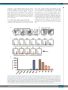
Figure 1. Tfr1 expression in hematopoietic stem/progenitor cells. (A) Representative gating strategy for analyzing long-term hematopoietic stem cells (HSC) (LT-HSC, Lin–cKit+Sca1hiCD34–Flt3–), short-term HSC (ST-HSC, Lin–cKit+Sca1hiCD34+Flt3–), multipotent progenitor (MPP, Lin–cKit+Sca1hiCD34+ Flt3+) cells, common myeloid pro- genitor (CMP, Lin–cKit+Sca1-CD34+ CD16/32int) cells, granulocyte-macrophage progenitor (GMP, Lin-cKit+Sca1-CD34+CD16/32hi) cells, megakaryocyte-erythroid pro- genitor (MEP, Lin–cKit+Sca1-CD34–CD16/32lo) cells, and common lymphoid progenitor (CLP, Lin–cKitintSca1intIL-7R+Flt3+) cells. (B) Erythrocytes gating strategy was used based on the expression levels of Ter119 and CD44: R1, proerythroblasts (Ter119loCD44hi); R2, early basophilic erythroblasts (CD44hiFSChi); R3, polychromatophilic erythroblasts (CD44hiFSCmed); R4, orthochromatophilic erythroblasts (CD44medFSCmed); and R5, mature erythrocytes (CD44loFSClo). (C) Flow cytometric analysis of Tfr1 expression in HSPC and erythroblasts in fetal liver of E16.5 mouse embryos. Grey histogram: isotype control (Rat IgG2a, k-PE). (D) Quantification of the flow cytometry data in C, showing the mean fluorescence intensity (MFI) of Tfr1 in the indicated hematopoietic stem/progenitor cells (HSPC) and erythroblasts.
haematologica | 2020; 105(8)
2073
