
Page 100 - 2020_08-Haematologica-web
P. 100
S. Wang et al.
A
B
CD
E
F
G
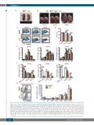
Figure 4. Loss of Tfr1 in hematopoietic stem cells causes progressively impaired cellular maturation in fetal liver. (A) Representative images of a control and Tfr1 knockout (cKO) embryo at P1. (B) Representative images of control and cKO embryos at E14.5, E16.5, and E18.5. (C) Representative flow cytometric profiles of Lin+ and Lin- cell populations in the liver of control and cKO embryos at E14.5, E16.5, and E18.5. (D) Percentage of Lin+ cells in the liver of control and cKO embryos at E14.5, E16.5, and E18.5 (n=5 per group). (E) Absolute numbers of the indicated erythrocyte development stages in the liver of control and cKO embryos at E14.5, E16.5, and E18.5 (n=5 per group). (F) Absolute number of myeloid cells (Mac1+Gr1+), B cells (CD19+), and T cells (CD3+) in the liver of control and cKO embryos at E14.5, E16.5, and E18.5 (n=5 per group). (G) Hematopoietic progenitor cells (HPC) were gated for fetal liver cells in control and cKO embryos using flow cytometry (left panel). The bar graph (right panel) shows the absolute numbers of LT, ST, MPP, CLP, CMP, GMP, and MEP cells in control and cKO fetal liver cells at E16.5 (n = 5 for each group); note the break in the y-axis. LT: long-term HSC; ST: short-term HSC; MPP: multipotent progenitor cells; CLP: common lymphoid progenitor cells; CMP: common myeloid progenitor cells; GMP: granulocyte/monocyte progenitor cells; MEP: megakaryocyte/erythrocyte progenitor cells. *P<0.05; **P<0.01; ***P<0.001.
2076
haematologica | 2020; 105(8)
