
Page 104 - 2020_08-Haematologica-web
P. 104
S. Wang et al.
Discussion
Tfr1 has long been used as a marker of red blood cells and is believed to play an essential role in erythropoiesis; however, its role in HSPC is poorly understood. Here, we generated and characterized a mouse model in which Tfr1 expression was deleted specifically in HSC and observed profoundly impaired BM function and defects in multiple cell lineages. These defects, which cause cKO offspring to die within one week of age, indicate that Tfr1 plays an essential role in hematopoiesis.
Specifically, our HSC-specific Tfr1-deficient mouse model allowed us to systematically dissect the role of Tfr1 in the development of erythrocytes, granulocytes, throm- bocytes, and lymphocytes. Our findings of microcytic hypochromic anemia in neonatal cKO pups and progressive erythropenia in FL of cKO embryos reveal that Tfr1 is required for erythropoiesis at an early stage, as loss of Tfr1 primarily blocked the differentiation of erythroblast precur- sors (e.g. proerythroblasts, polychromatophilic erythrob- lasts), leading to decreased mature erythrocytes. In addi- tion, although T-cell development was severely impaired in cKO mice, B-cell development was largely unaffected. These findings are supported by previous experiments showing that developing T cells derived from Tfr1-/- ES cells arrested in an early stage, whereas B-cell development was affected less severely.14 Moreover, a recent study by Wang et al. found that Tfr1fl/fl;Cd4-Cre mice develop normally, but have reduced production of pro-inflammatory cytokines.30
Although the production of B cells was generally unaffected in our cKO mice, antibody production was likely affected given that patients with a mutation in the TFR1 gene have severe hypogammaglobulinemia.15 Importantly, we found that monocyte development is severely blocked in embry- onic development in cKO mice, providing the first direct evidence that Tfr1 plays an important role in monocyte development, which is consistent with studies demonstrat- ing the role of Tfr1 in monocytes.31-33
A wide range of peripheral blood cell types were decreased in cKO mouse, while the number of platelets was increased. On one hand, iron deficiency anemia has been shown to cause reactive thrombocytosis in both patients34 and animal models,35 and iron deficiency itself can promote megakaryopoiesis.36 On the other hand, megakaryocytes are reported to arise directly from stem- like megakaryocyte-committed progenitor cells, a popula- tion that shares many features with multipotent HSC and serves as a megakaryocyte lineage-restricted emergency pool,37 these progenitor cells might escape the impaired proliferation that affects other cell types.
Although, Tfr1fl/fl;Vav-Cre mice have concomitant dele- tion of Tfr1 in hematopoietic and endothelial cells, it remains elusive with respect to the effect of endothelial Tfr1 on systemic iron regulation. Most recently, Canali et al. and Koch et al. independently reported that liver sinu- soidal endothelial cells (LSEC) are the major source of bone morphogenetic proteins (BMP), which are essential to the hepcidin expression.38,39 Specifically, it is suggested
A
B
C
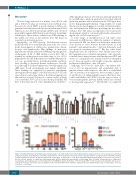
Figure 7. Treating Tfr1 knockout hematopoietic stem/progenitor cells with hemin restores their differentiation capacity. (A) In vivo colony assays were performed using hematopoietic stem/progenitor cells (HSPC) obtained from control and Tfr1 knockout (cKO) embryos at E14.5 (n=3 for each group) and treated with or without 5 mM holo-transferrin (holo-Tf), 10 mM ferric ammonium citrate (FAC), 10 mM hemin, or 0.25 mM Fe (III)/8-hydroxyquinoline (Fe/8-HQ). Colonies were measured on day 12, and the number of erythrocyte burst-forming units (BFU-E), granulocyte/macrophage colony-forming units (CFU-GM), and granulocytes, erythrocytes, macrophages, and megakaryocyte colony-forming units (CFU-GEMM) is shown. (B) Number of CFU formed by HSPC of cKO fetal liver treated with the indicated con- centrations of hemin, and control cells. (C) Number of CFU formed by HSPC infected with the indicated Tfr1-expressing lentivirus. N.D.: not detectable. *P<0.05; **P<0.01; ***P<0.001.
2080
haematologica | 2020; 105(8)
