
Page 103 - 2020_08-Haematologica-web
P. 103
The critical function of Tfr1 in hematopoiesis
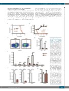
Overexpressing wild-type Tfr1 but not the R654A mutant rescues Tfr1-deficient HSPC
Noncanonical functions of Tfr1 have been reported, including maintaining intestinal epithelial function inde- pendent of iron uptake,16 cell survival,27 and regulating mitochondrial morphology.28 Additionally, the missense mutation R654A in Tfr1 prevents Tf binding without affecting the receptor’s ability to bind Hfe protein.29 We therefore expressed either wild-type Tfr1 or mutant Tfr1 in cKO FL cells using lentivirus infection. Expressing wild-
AB
P=0.0021 CD
E
type Tfr1 in cKO cells rescued the colony-forming capac- ity; interestingly, expressing the Tfr1L622A mutant (which lacks the ability to bind Hfe) partially rescued, whereas expressing the Tfr1R654A mutant was completely unable to rescue the impaired colony-forming capacity of cKO cells (Figure 7C). These results suggest that its function in iron uptake rather than signal transduction is the sole mecha- nism by which Tfr1 controls differentiation and survival in the hematopoietic system.
Figure 6. Tfr1 knockout hemtopoi- etic stem cells have impaired
F
G and CLP (common lymphoid pro- genitor cells) were measured 16 weeks after co-transplantation with control and cKO donor cells (n=3 for each group). (F) The absolute number of donor-derived myeloid cells (Mac1+Gr1+) and B cells (B220+) in the spleen, and T cells (CD3+) in the thymus of recip- ient mice 16 weeks after co-trans- plantation with control and cKO cells (n=3 for each group). (G) Homing efficiency of fetal liver cells obtained from either control or cKO mice 40 hours after transplanta- tion into lethally irradiated recipi- ent mice (n=3 per group). *P<0.05; **P<0.01; ***P<0.001.
capacity for
hematopoiesis following trans- plantation. (A) Kaplan–Meier sur- vival curve of CD45.1 mice that were lethally irradiated and then transplanted with fetal liver cells obtained from CD45.2 control or Tfr1 knockout (cKO) embryos (n=5 recipients per group). (B) Recipient mice received a 50/50 combina- tion of the indicated donor-derived fetal liver (CD45.2) cells and com- petitor (CD45.1) cells were meas- ured at the indicated time points following transplantation (n=3 recipients per group). (C) Donor- derived (CD45.2+) lineage-negative cells (Lin–) and mature hematopoi- etic cells (Lin+) were gated by flow cytometry. (D) The percentage of donor-derived Lin– and Lin+ cells in the recipient bone marrow cells after transplantation for 16 weeks. (E) The absolute number of donor- derived LT (long-term HSC), ST (short-term HSC), MPP (multipo- tent progenitor cells), CMP (com- mon myeloid progenitor cells), GMP (granulocyte/monocyte pro- genitor cells), MEP (megakary- ocyte/erythrocyte progenitor cells),
restoring
haematologica | 2020; 105(8)
2079
