
Page 157 - 2020_07-Haematologica-web
P. 157
Genetic alterations in indolent GI T-cell LPD
cases and in the CD4-/CD8- case. CD103 expression was detected in two of four CD8+ cases (Figure 2H), with one also showing partial CD56 expression (case 8) (Figure 2I). The CD4+/CD8+ and the CD4-/CD8- cases expressed PD- 1. CD20 highlighted mucosal lymphoid follicles, but the neoplastic cells were CD20- in all ITLPD. Surface TCRαβ expression was observed in all cases evaluated by flow cytometry and none expressed TCRgd. All analyzed cases were negative for BCL6, CD10, FoxP3, MATK, PD- L1 or CD30, however CD30 expression (and acquisition of cytotoxic proteins) was observed, and previously reported, upon large cell transformation (case 4).11 The Ki-67 proliferation index was low (<5%) in all ITLPD evaluated (Figures 1J and 2J).
Determination of the cell of origin
Since a good correlation between the transcriptional profiles and immunohistochemistry for T-bet and GATA3 has been reported in T-cell lymphomas,27 we assessed T- bet and GATA3 expression by immunohistochemistry to determine the cell of origin of ITLPD (Table 2, Online Supplementary Table S1, Online Supplementary Figure S1). The CD4+ cases showed heterogeneity with regards to T-
bet and GATA3 expression: one case each was T-bet+ and GATA3+, suggesting T-helper type 1 (Th1) and type 2 (Th2) lineage, respectively and two cases showed T-bet and GATA3 co-expression - hybrid Th1/Th2 profile (Figure 1K, L). The CD4+/CD8+ ITLPD also co-expressed T-bet and GATA3. The CD4-/CD8- case and three of the four (75%) CD8+ cases were GATA3+, implying a type-2- polarized effector T-cell (Tc2) phenotype and one CD8+ case showed T-bet and GATA3 co-expression (Figure 2K, L). Sequential analysis of one CD4+ ITLPD (case 2) showed a shift from a Th1/Th2 (T-bet and GATA3 co- expression) to Th2 (GATA3) phenotype over the course of disease. Double staining for T-bet and GATA3, performed in a subset (cases 2, 7, and 8), confirmed distinct T-bet and GATA3+ as well as T-bet and GATA3 co-expressing lym- phocytes (data not shown).
T-cell receptor gene rearrangement analysis
Clonal TRB and/or TRG rearrangement products were detected in all ITLPD. In patients in whom longitudinal testing was performed, similar sized peaks were observed in all samples, confirming persistence of the same lym- phocytic clone.
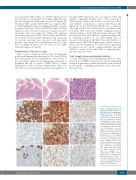
ABC
DEF
GHI
Figure 2. Morphological and immunophenotypic features of CD8+ indolent T-cell lymphopro- liferative disorders of the gas- trointestinal tract. (A) An ileal biopsy (case 8) shows a dense mucosal lymphocytic infiltrate expanding the lamina propria and widening the villi; no villous atrophy is present but the crypts are hyperplastic. (B) Small clus- ters of lymphocytes are seen within the villus epithelium along the lateral edges. There is no increase in intraepithelial lymphocytes. (C) The lympho- cytes are small and have round or oval nuclei, condensed chro- matin, indistinct nucleoli, and scant to moderate clear or pale pink cytoplasm. The lympho- cytes express (D) CD8 and (E)
and (G) granzyme B expressed by a subset. (H) The lymphocytes are CD103+ and a subset expresses (I) CD56. (J) The Ki-67 proliferation index is low (<5%). The majority of cells express (K) GATA3, but 60% also show (L) T-bet expression.
JKL CD3. Most of the cells express the cytotoxic marker (F) TIA1
is
haematologica | 2020; 105(7)
1899
