
Page 232 - Haematologica - Vol. 105 n. 6 - June 2020
P. 232
C. Han et al.
pcEV from injured placenta could induce a preeclampsia- like phenotype in pregnant mice.
Placenta-derived extracellular vesicles directly induced hypertension and proteinuria
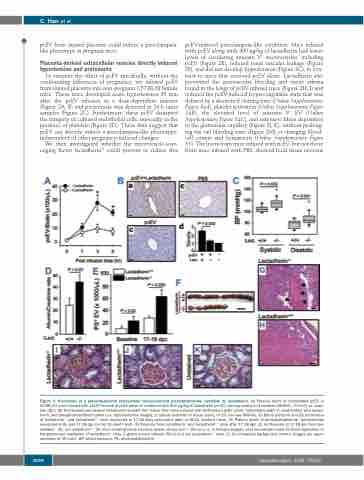
To examine the effect of pcEV specifically, without the confounding influences of pregnancy, we infused pcEV from injured placenta into non-pregnant C57 BL/6J female mice. These mice developed acute hypertension 30 min after the pcEV infusion in a dose-dependent manner (Figure 2A, B) and proteinuria was detected in 24-h urine samples (Figure 2C). Furthermore, these pcEV disrupted the integrity of cultured endothelial cells, especially in the presence of platelets (Figure 2D). These data suggest that pcEV can directly induce a preeclampsia-like phenotype, independent of other pregnancy-induced changes.
We then investigated whether the microvesicle-scav- enging factor lactadherin28 could prevent or reduce this
pcEV-induced preeclampsia-like condition. Mice infused with pcEV along with 400 μg/kg of lactadherin had lower levels of circulating annexin V+ microvesicles, including pcEV (Figure 2E), reduced renal vascular leakage (Figure 2F), and did not develop hypertension (Figure 2G), in con- trast to mice that received pcEV alone. Lactadherin also prevented the perivascular bleeding and tissue edema found in the lungs of pcEV-infused mice (Figure 2H, I) and reduced the pcEV-induced hypercoagulable state that was defined by a shortened clotting time (Online Supplementary Figure S4A), platelet activation (Online Supplementary Figure S4B), the elevated level of annexin V+ EV (Online Supplementary Figure S4C), and extensive fibrin deposition in the glomerular capillary (Figure 2J, K), without prolong- ing the tail bleeding time (Figure 2M) or changing blood- cell counts and hematocrit (Online Supplementary Figure S5). The livers from mice infused with pcEV, but not those from mice infused with PBS, showed focal tissue necrosis
Figure 3. Prevention of a placenta-derived extracellular vesicle-induced preeclampsia-like condition by lactadherin. (A) Plasma levels of biotinylated pcEV in C57BL/6J mice infused with 1x10⁷/mouse of pcEV alone or combined with 400 μg/kg of lactadherin [n=10, one-way analysis of variance (ANOVA), *P<0.01 vs. base- line (BL)]. (B) Horseradish peroxidase streptavidin-stained liver tissue from mice infused with biotinylated pcEV alone, biotinylated pcEV in combination with lactad- herin, and phosphate-buffered saline (a-c: representative images, d: optical densities of tissue scans, n=19, one-way ANOVA). (C) Blood pressure and (D) proteinuria of lactadherin-/- and lactadherin+/+ mice measured at 17-18 days post-coitus (dpc) (n=8-24, Student t-test). (E) Plasma levels of phosphatidylserine+ microvesicles measured at BL and 17-18 dpc (n=16, Student t-test). (F) Placenta from lactadherin-/- and lactadherin+/+ mice after 17-18 dpc. (G, H) Placenta at 17-18 dpc from lac- tadherin-/- (G), not lactadherin+/+ (H) mice showing tissue necrosis (black arrows, bar = 100 μm). (I, J) Phosphotungstic acid hematoxylin stain for fibrin deposition in the glomerular capillaries of lactadherin-/- mice (I, green arrows indicate fibrin) and not lactadherin+/+ mice (J). (K) Unstained background control. Images are repre- sentative of 39 mice. BP: blood pressure; PS: phosphatidylserine.
1690
haematologica | 2020; 105(6)
