
Page 233 - Haematologica - Vol. 105 n. 6 - June 2020
P. 233
Placental extracellular vesicles induce preeclampsia
and infiltration by inflammatory cells; these changes were not detected in pcEV-infused mice that also received lac- tadherin (Online Supplementary Figure S6). In contrast to kidney tissues, intravascular fibrin deposition was very limited in the livers of pcEV-infused mice.
To measure directly whether lactadherin enhanced pcEV clearance, biotinylated pcEV (1×10⁷/mouse) were infused into non-pregnant C57BL/6J mice along with 400 μg/kg of lactadherin or an equal volume of PBS. The plas- ma level of biotinylated pcEV reached a plateau 3 h after infusion (Figure 3A) but was significantly lower in mice that also received lactadherin (Figure 3A). The mice given pcEV and lactadherin had more extensive accumulation of biotinylated pcEV in their livers (Figure 3B).
Lactadherin-deficient mice developed unprovoked preeclampsia during pregnancy
To further validate the role of lactadherin in promoting EV clearance and preventing preeclampsia, we also exam-
ined lactadherin-/- mice and their wildtype littermates (Online Supplementary Figure S7). At 17-18 dpc, lactadherin-/- mice had higher BP (Figure 3C) and developed proteinuria (Figure 3D) without pcEV infusion, as required for pregnant C57BL/6J mice (Figure 1H). Plasma levels of PS-expressing EV recognized by annexin V were significantly higher in lactadherin-/- mice than in their wildtype littermates at base- line, and they increased further during pregnancy (Figure 3E). There was no visible difference in the appearance of the fetuses and sizes of placenta between lactadherin-/- and lactadherin+/+ mice at 17-18 dpc (Online Supplement Figure S8), but the number of lactadherin-/- litters was significantly less than the number of lactadherin+/+ litters (8.2±1.7 vs. 11.5±1.4, Student t-test, P<0.05) (Figure 3F). Hematoxylin & eosin stains of placental tissues frequently detected tissue necrosis in the trophoblast villi and decidua of lactadherin-/- mice (Figure 3G,H). The lactadherin deficiency also resulted in more extensive fibrin deposition in glomerular capillaries, which occurred less in wildtype littermates (Figure 3I-K).
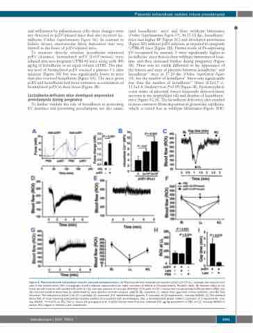
Figure 4. Placenta-derived extracellular vesicles induced vasoconstriction. (A) Placenta-derived extracellular vesicles (pcEV) (2×10⁵/μL) increase the vascular ten- sion of the carotid artery (left: a myograph of pcEV-induced vasoconstriction; right: summary of effects in 20 experiments, Student t-test). (B) Calcium influx of cul- tured smooth muscle cells treated with pcEV [n=12, one-way analysis of variance (ANOVA)]. (C-F) pcEV (1×10⁷/mouse) but not phosphate-buffered saline (PBS) rap- idly reduced cerebral blood flow as determined by laser speckle contrast analysis (LASCA) (BL: baseline; LL: lowest level, gap-time of flow reduction; and RC: flow recovery). The reduction is either fully (C) or partially (D) recovered (C-E: representatives graphs; F: summary of 12 experiments, one-way ANOVA). (G) The cerebral blood flow of mice receiving extracellular vesicles purified from patients with preeclampsia (top: a representative graph, bottom: summary of 3 experiments, one- way ANOVA, *P<0.001 vs. BL). The LL values (H) and gap-time (I) of pcEV-infused mice that also received 400 μg/kg lactadherin or PBS (n=12, one-way ANOVA on ranks). ROI: region of interest; Lact: lactadherin.
haematologica | 2020; 105(6)
1691
