
Page 231 - Haematologica - Vol. 105 n. 6 - June 2020
P. 231
Placental extracellular vesicles induce preeclampsia
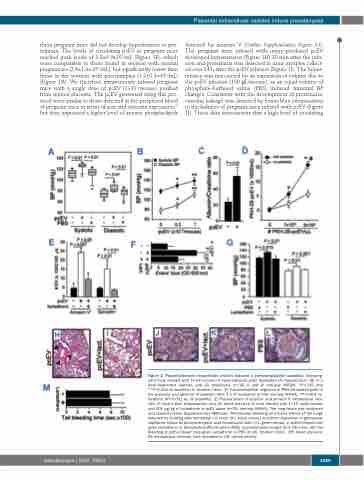
these pregnant mice did not develop hypertension or pro- teinuria. The levels of circulating pcEV in pregnant mice reached peak levels of 3.8±0.9x10⁷/mL (Figure 1F), which were comparable to those found in women with normal pregnancies (2.6±1.3×10⁷/mL), but significantly lower than those in the women with preeclampsia (1.2±0.3×10⁸/mL) (Figure 1A). We therefore intravenously infused pregnant mice with a single dose of pcEV (1×10⁷/mouse) purified from injured placenta. The pcEV generated using this pro- tocol were similar to those detected in the peripheral blood of pregnant mice in terms of size and syncytin expression,31 but they expressed a higher level of anionic phospholipids
detected by annexin V (Online Supplementary Figure S3). The pregnant mice infused with injury-produced pcEV developed hypertension (Figure 1H) 30 min after the infu- sion and proteinuria was detected in urine samples collect- ed over 24 h after the pcEV infusion (Figure 1I). The hyper- tension was not caused by an expansion of volume due to the pcEV infusion (100 μL/mouse), as an equal volume of phosphate-buffered saline (PBS) induced minimal BP changes. Consistent with the development of proteinuria, vascular leakage was detected by Evans blue extravasation in the kidneys of pregnant mice infused with pcEV (Figure 1J). These data demonstrate that a high level of circulating
Figure 2. Placenta-derived extracellular vesicles induced a preeclampsia-like condition. Non-preg- nant mice infused with 1×10⁷/mouse of injury-released pcEV developed (A) hypertension, (B) in a dose-dependent manner, and (C) proteinuria (n=18, A and B: one-way ANOVA, *P<0.05 and **P<0.001 vs. baseline; C: Student t-test). (D) Transendothelial migration of PKH-26-labeled pcEV in the presence and absence of platelets after 3 h of incubation (n=26, one-way ANOVA, *P<0.001 vs. baseline, #P<0.001 vs. no platelets). (E) Plasma levels of syncytin+ and annexin V+ extracellular vesi- cles, (F) Evans blue extravasation, and (G) blood pressure of mice infused with 1×10⁷ pcEV/mouse and 400 μg/kg of lactadherin or pcEV alone (n=26, one-way ANOVA). The lung tissue was sectioned and stained (Online Supplementary Methods). Perivascular bleeding and tissue edema of the lungs detected by staining with hematoxylin & eosin (H-I, black arrows) and fibrin deposition in glomerular capillaries shown by phosphotungstic acid hematoxylin stain (J-L, green arrows) in pcEV-infused mice given lactadherin or phosphate-buffered saline (PBS) (representative images from 26 mice). (M) Tail bleeding of pcEV-infused mice given lactadherin or PBS (n=26, Student t-test). BP: blood pressure; EV: extracellular vesicles; Lact: lactadherin; OD: optical density.
haematologica | 2020; 105(6)
1689
