
Page 230 - Haematologica - Vol. 105 n. 6 - June 2020
P. 230
C. Han et al.
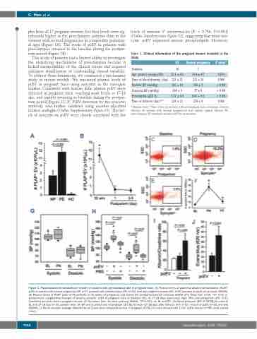
ples from all 17 pregnant women, but their levels were sig- nificantly higher in the preeclamptic patients than in the women with normal pregnancies at comparable gestation- al ages (Figure 1A). The levels of pcEV in patients with preeclampsia returned to the baseline during the postpar- tum period (Figure 1B).
This study of patients had a limited ability to investigate the underlying mechanisms of preeclampsia because it lacked manipulability of the clinical course and required extensive stratification of confounding clinical variables. To address these limitations, we conducted a mechanistic study in mouse models. We measured plasma levels of pcEV in pregnant mice using syncytin as the surrogate marker. Consistent with human data, plasma pcEV were detected in pregnant mice, reaching peak levels at 17-18 dpc, and rapidly returning to baseline during the postpar- tum period (Figure 1C-F). PcEV detection by the syncytin antibody was further validated using another placental marker, endoglin (Online Supplementary Figure S1). The lev- els of syncytin on pcEV were closely correlated with the
levels of annexin V+ microvesicles (R2 = 0.766, P<0.001) (Online Supplementary Figure S2), suggesting that most syn- cytin+ pcEV expressed anionic phospholipids. However,
Table 1. Clinical information of the pregnant women included in the study.
PE
Number 10
Normal pregnancy
7
31.4 ± 4.7 233 ± 20 122 ± 7 77 ± 8 0.01 + 0.0 270 ± 9
P value* 0.219
0.989 < 0.001 < 0.001 < 0.001 0.003
Age (years) (mean±SD) Time of blood drawing (day) Systolic BP (mmHg) Diastolic BP (mmHg) Proteinuria (g/24 h)
Time of delivery (day)**
33.1 ± 4.2 233 ± 21 166 ± 14 104 ± 9 5.37 ± 4.4 239 ± 23
*Student t-test; **Nine of the ten women with preeclampsia had a Cesarean delivery, whereas all women with normal pregnancies had natural vaginal delivery. PE: preeclampsia; SD: standard deviation; BP: blood pressure.
Figure 1. Placenta-derived extracellular vesicles in women with preeclampsia and in pregnant mice. (A) Plasma levels of placental alkaline phosphatase (PLAP)+ pcEV in women with normal pregnancy (NP, n=7), patients with preeclampsia (PE, n=10), and non-pregnant women (HC, n=5) (one-way analysis of variance, ANOVA). (B) Plasma levels of PLAP+ pcEV of PE patients at 32 weeks of pregnancy and during the postpartum period (one-way ANOVA with Tukey test, n=10, *P< 0.01 vs. postpartum). Longitudinal changes of plasma syncytin+ pcEV of pregnant mice at baseline (BL), at 17-18 days post-coitus (dpc) (PN) and postpartum (PT). (C-E): Cytometry dot plots from a pregnant mouse. (F) Summary from 15 mice (one-way ANOVA, *P<0.001, vs. BL and PT). (G) Blood pressure (BP) of C57BL/6J mice at BL and 17-18 dpc (n=30, paired t test). (H) BP and (I) proteinuria of pregnant C57 BL/6J mice (17-18 dpc) after infusion with 1×10⁷/mouse of pcEV (n=32, one-way ANOVA). (J) Renal vascular leakage detected by an Evans blue extravasation test in pregnant C57BL/6J mice infused with 1×10⁷ pcEV/mouse or PBS (n=8, paired t test).
1688
haematologica | 2020; 105(6)
