
Page 222 - Haematologica - Vol. 105 n. 6 - June 2020
P. 222
C. Kroone et al.
the ligated arteries in five of 14 FHL2-KO mice, whereas such structures were absent in the vascular lesions of WT mice (data not shown). We therefore hypothesized that FHL2 may be involved in thrombus formation. To sub- stantiate this hypothesis, we performed FeCl3-induced thrombosis experiments in WT and FHL2-KO mice. Upon
FeCl3-induced vascular injury, the blood flow in mesen- teric veins was imaged by intravital microscopy to assess thrombus formation. Platelets were visualized with an anti-GPIbβ antibody and fluorescent fibrinogen was injected to monitor fibrin accumulation (Figure 1A). The time from vessel injury to formation of a stable occlusive
A
BCE
D
FG
H
IJ
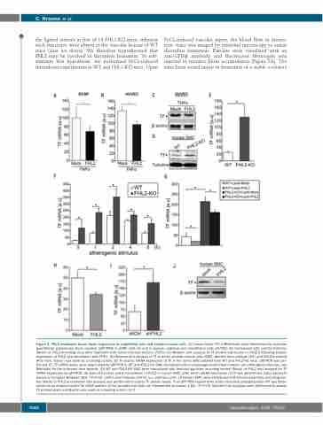
Figure 2. FHL2 modulates tissue factor expression in endothelial cells and smooth muscle cells. (A) Tissue factor (TF) mRNA levels were determined by real-time quantitative polymerase chain reaction (qRT-PCR) in ECRF cells (A) and in human umbilical vein endothelial cells (HUVEC) (B) transduced with control lentivirus (Mock) or FHL2-encoding virus after treatment with tumor necrosis factor-α (TNFα) (C) Western blot analysis of TF protein expression in HUVEC following ectopic expression of FHL2 and stimulation with TNFα. (D) Western blot analysis of TF in aortic smooth muscle cells (SMC) derived from wildtype (WT) and FHL2-knockout (KO) mice. Tubulin was used as a loading control. (E) To assess mRNA expression of TF in the aortic SMC isolated from WT and FHL2-KO mice, qRT-PCR was per- formed. (F) TF mRNA levels were determined by qRT-PCR in WT and FHL2-KO SMC stimulated with macrophage-conditioned medium (an atherogenic stimulus, see Methods) for the indicated time periods. (G) WT and FHL2-KO SMC were transduced with lentiviral particles encoding control (Mock) or FHL2 and assayed for TF mRNA expression by qRT-PCR. (H) Gain-of-function and (I) knockdown of FHL2 in human SMC, after which mRNA expression of TF was determined. Data represent means ± standard deviation (SD). *P<0.05. shCtrl, short-hairpin control. a.u, arbitrary units. (J) Human SMC were transduced with lentiviral particles encoding con- trol (Mock) or FHL2 and western blot analysis was performed to assess TF protein levels. In all qRT-PCR experiments acidic ribosomal phosphoprotein P0 was deter- mined as an internal control for cDNA content of the samples and data are represented as means ± SD. *P<0.05. Western blot analyses were performed to assess TF protein levels and β-actin was used as a loading control. n=3
1680
haematologica | 2020; 105(6)
