
Page 178 - Haematologica - Vol. 105 n. 6 - June 2020
P. 178
J. Tong et al.
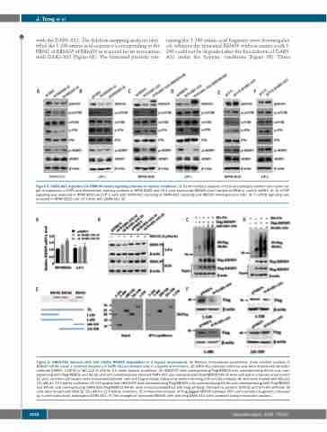
with the DARS-AS1. The deletion-mapping analyses iden- tified the 1-240 amino acid sequence (corresponding to the RRM1 of RBM39) of RBM39 as required for its association with DARS-AS1 (Figure 6E). The truncated proteins con-
taining the 1-240 amino acid fragment were downregulat- ed, whereas the truncated RBM39 without amino acids 1- 240 could not be degraded after the knockdown of DARS- AS1 under the hypoxic conditions (Figure 6F). These
ABCDEF
Figure 5. DARS-AS1 regulates the RBM39-related signaling pathway in hypoxic conditions. (A, B) Immunoblot analysis of total and phosphorylated mammalian tar- get of rapamycin (mTOR) and downstream pathway proteins in RPMI 8226 and LP-1 cells expressing RBM39 short hairpin (sh)RNA or control shRNA. (C, D) mTOR signaling was analyzed in RPMI 8226 and LP-1 cells with DARS-AS1 silencing or DARS-AS1 silencing and RBM39 overexpression (OE). (E, F) mTOR signaling was analyzed in RPMI 8226 and LP-1 cells with DARS-AS1 OE.
ABCD
EF
Figure 6. DARS-AS1 interacts with and inhibits RBM39 degradation in a hypoxic environment. (A) Reverse transcriptase polymerase chain reaction analysis of RBM39 mRNA (mean ± standard deviation) in DARS-AS1 knockdown cells in a hypoxic environment. (B) DARS-AS1-silenced myeloma cells were treated with dimethyl- sulfoxide (DMSO, 1:1000) or MG-132 (5 μM) for 6 h under hypoxic conditions. (C) HEK293T cells overexpressing Flag-RBM39 only, overexpressing HA-Ub only, over- expressing both Flag-RBM39 and HA-Ub, and with simultaneously silenced DARS-AS1 and overexpressed Flag-RBM39/HA-Ub were cultured in a hypoxic environment for 24 h, and then cell lysates were immunoprecipitated with anti-Flag antibody, followed by western blotting with anti-HA antibody. All cells were treated with MG132 (10 μM) for 12 h before collection. (D) Cell lysates from HEK293T cells overexpressing Flag-RBM39 only, overexpressing HA-Ub only, overexpressing both Flag-RBM39 and HA-Ub, and overexpressing DARS-AS1/Flag-RBM39/HA-Ub were immunoprecipitated with Flag antibody, followed by western blotting with anti-HA antibody. All cells were treated with MG132 (10 μM) for 12 h before collection. (E) Immunoblot analysis of Flag-tagged RBM39 [wildtype (WT) and truncation fragments] retrieved by in vitro-transcribed, biotinylated DARS-AS1. (F) The changes of truncated RBM39 after silencing DARS-AS1 were assessed using immunoblot analysis.
1636
haematologica | 2020; 105(6)
