
Page 179 - Haematologica - Vol. 105 n. 6 - June 2020
P. 179
DARS-AS1 promotes myeloma malignancy in hypoxia
results suggest that the 1–240 amino acid sequence of RBM39 is essential for its binding to DARS-AS1.
The interaction of DARS-AS1 with RBM39 inhibits the degradation of RBM39 through RNF147
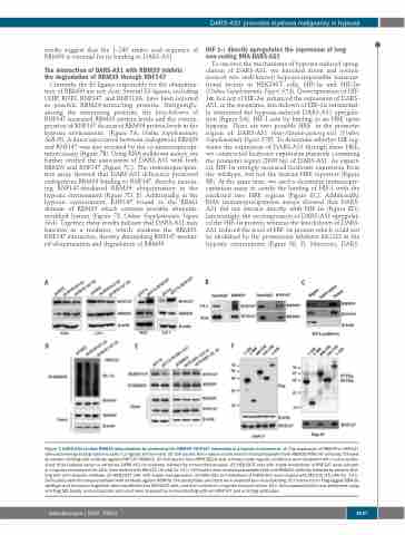
Currently, the E3 ligases responsible for the ubiquitina- tion of RBM39 are not clear. Several E3 ligases, including CHIP, RNF2, RNF147, and RNF113A, have been reported as possible RBM39-interacting proteins. Intriguingly, among the interacting proteins, the knockdown of RNF147 increased RBM39 protein levels and the overex- pression of RNF147 decreased RBM39 protein levels in the hypoxic environment (Figure 7A, Online Supplementary S6B-D). A direct association between endogenous RBM39 and RNF147 was also revealed by the co-immunoprecipi- tation assays (Figure 7B). Using RNA pulldown assays, we further verified the association of DARS-AS1 with both RBM39 and RNF147 (Figure 7C). The immunoprecipita- tion assay showed that DARS-AS1 deficiency promoted endogenous RBM39 binding to RNF147, thereby increas- ing RNF147-mediated RBM39 ubiquitination in the hypoxic environment (Figure 7D, E). Additionally, in the hypoxic environment, RNF147 bound to the RRM1 domain of RBM39 which contains possible ubiquitin- modified lysines (Figure 7F, Online Supplementary Figure S6A). Together, these results indicate that DARS-AS1 may function as a mediator, which weakens the RBM39- RNF147 interaction, thereby diminishing RNF147-mediat- ed ubiquitination and degradation of RBM39.
HIF-1α directly upregulates the expression of long non-coding RNA-DARS-AS1
To uncover the mechanisms of hypoxia-induced upreg- ulation of DARS-AS1, we knocked down and overex- pressed two well-known hypoxia-responsible transcrip- tional factors in HEK293T cells, HIF-1α and HIF-2α (Online Supplementary Figure S7A). Overexpression of HIF- 1α, but not of HIF-2α, enhanced the expression of DARS- AS1; in the meantime, knockdown of HIF-1α substantial- ly attenuated the hypoxia-induced DARS-AS1 upregula- tion (Figure 8A). HIF-1 acts by binding to an HRE upon hypoxia. There are two possible HRE in the promoter region of DARS-AS1 (http://jaspar.genereg.net) (Online Supplementary Figure S7B). To determine whether HIF reg- ulates the expression of DARS-AS1 through these HRE, we constructed luciferase expression plasmids containing the promoter region (2000 bp) of DARS-AS1. As expect- ed, HIF-1α strongly increased luciferase expression from the wildtype, but not the mutant HRE reporters (Figure 8B). At the same time, we used a chromatin immunopre- cipitation assay to certify the binding of HIF-1 with the predicted two HRE regions (Figure 8C). Additionally, RNA immunoprecipitation assays showed that DARS- AS1 did not interact directly with HIF-1α (Figure 8D). Interestingly, the overexpression of DARS-AS1 upregulat- ed the HIF-1α protein, whereas the knockdown of DARS- AS1 reduced the level of HIF-1α protein which could not be abolished by the proteasome inhibitor MG132 in the hypoxic environment (Figure 8E, F). Moreover, DARS-
ABC
DEF
Figure 7. DARS-AS1 inhibits RBM39 ubiquitination by weakening the RBM39–RNF147 interaction in a hypoxic environment. (A) The expression of RBM39 in RNF147 silenced/overexpressing myeloma cells in a hypoxic environment. (B) Cell lysates from myeloma cells were immunoprecipitated with RBM39/RNF147 antibody, followed by western blotting with antibody against RNF147/RBM39. (C) Cell lysates from RPMI 8226 cells cultured under hypoxic conditions were incubated with in vitro-synthe- sized, biotin-labeled sense or antisense DARS-AS1 for pulldown followed by immunoblot analysis. (D) HEK293T cells with stable knockdown of RNF147 were cultured in a hypoxic environment for 24 h, then treated with MG132 (10 μM) for 12 h. Cell lysates were immunoprecipitated with anti-RBM39 antibody, followed by western blot- ting with anti-ubiquitin antibody. (E) HEK293T cells with stable overexpression of DARS-AS1 or knockdown of DARS-AS1 were treated with MG132 (10 μM) for 12 h. Cell lysates were immunoprecipitated with antibody against RBM39. The precipitates and input were analyzed by immunoblotting. (F) Constructs for Flag-tagged RBM39 (wildtype and truncation fragment) were transfected into HEK293T cells, and then cultured in a hypoxic environment for 24 h. Immunoprecipitation was performed using anti-Flag M2 beads, and precipitates and input were analyzed by immunoblotting with anti-RNF147 and anti-Flag antibodies.
haematologica | 2020; 105(6)
1637
