
Page 175 - Haematologica - Vol. 105 n. 6 - June 2020
P. 175
DARS-AS1 promotes myeloma malignancy in hypoxia
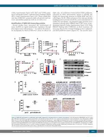
Online Supplementary Figure S3D). Ki67 and TUNEL stain- ing of xenografts further confirmed these findings (Figure 2H, J, Online Supplementary Figure S3F). These results indi- cate that DARS-AS1 promotes MM cell growth and sur- vival, and inhibits apoptosis both in vitro and in vivo.
Identification of DARS-AS1-interacting proteins
Recent studies have uncovered the involvement of many lncRNA in signal transduction regulation through their interactions with proteins.14,15 To explore the molecu- lar mechanism by which DARS-AS1 exerts its effects on
MM cells, we performed biotin-labeled RNA pulldown, followed by mass spectrometry assays to identify the DARS-AS1-interacting proteins in RPMI 8226 and LP-1 cells (Figure 3A, B). After exclusion of the non-specifically binding proteins present in both the sense and antisense of DARS-AS1 precipitate, six potential target proteins were identified, among which RBM39 and PCBP1 were of pre- dominant interest (Online Supplementary Figure S5A). Only RBM39 binding with DARS-AS1 in three myeloma cell lines was detected by western blotting in three independ- ent RNA pulldown assays (Figure 3C). The specific inter-
ABC
DEF
GH
IJ
Figure 2. DARS-AS1 participates in proliferation and inhibits apoptosis of myeloma cells in a hypoxic environment. (A, B) Cell growth of RPMI 8226 and LP-1 cells with DARS-AS1 knockdown in a hypoxic environment was determined every 24 h for 72 h using an automated cell counter (Countstar). (C, D) Cell growth of RPMI 8226 and LP-1 cells with DARS-AS1 overexpression was determined every day for 7 days using an automated cell counter (Countstar). (E) The rates of annexin V+ cells in myeloma cells with DARS-AS1 knockdown and control cells were determined by flow cytometry. (F) Cleaved caspase-3 and cleaved PARP were demonstrated by western blotting of RPMI 8226 and LP-1 cells with DARS-AS1 knockdown. (G, I) Xenograft mouse models of myeloma cells with DARS-AS1 overexpression or DARS- AS1 knockdown in NOD-SCID mice. (H, J) Ki67 staining of xenografts of DARS-AS1 overexpressing or DARS-AS1 knockdown cells.*P<0.05 and **P<0.01, compared with the control. Data are presented as the mean ± standard error of mean from three experiments.
haematologica | 2020; 105(6)
1633
