
Page 120 - Haematologica - Vol. 105 n. 6 - June 2020
P. 120
A. Touzart et al.
(ETP-ALL) lacked a significant distinct methylation signa- ture compared to non-ETP-ALL (Figure 3E).
Low level of promoter methylation predicted a poor outcome subgroup of adult T-ALL
T-ALL patients with the lowest methylation level (Q1, n=42/168) were significantly more men, were younger, and had a higher white blood cell (WBC) count at diagno- sis than patients with higher methylation levels (Table 1). Moreover, hypoM T-ALL demonstrated a significantly more frequent mature phenotype (TCRαβ+) and were associated with SIL-TAL1 rearrangement. They were also
significantly associated with a low rate of NOTCH1 path- way mutations and a high risk NOTCH1/FBXW7/RAS/ PTEN molecular classifier.22 In detail, we observed a signif- icantly lower incidence of NOTCH1/FBXW7 mutations and also a greater incidence of PTEN alterations (mutation and/or deletion) in the hypoM subgroup (CIMP-neg) as compared to the Int/High methylated cases (Online Supplementary Table S5). Despite a better bone marrow response at D8 (M1 status) in patients with low methyla- tion, we did not observe any impact of methylation on complete remission (CR) rate or post-induction minimal residual disease (MRD) level. In univariate analysis,
AB
C
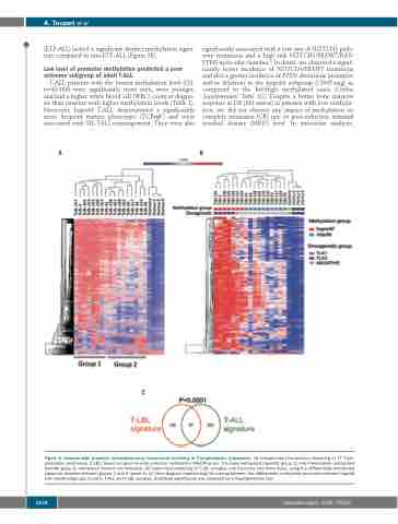
Figure 2. Genome-wide promoter methylation-array hierarchical clustering in T-lymphoblastic lymphomas. (A) Unsupervised hierarchical clustering of 17 T-lym- phoblastic lymphomas (T-LBL) based on genome-wide promoter methylation (MeDIP-array). The hypermethylated (hyperM; group 1) and intermediate methylated (interM; group 2) methylated clusters are indicated. (B) Supervised clustering of T-LBL samples, one thymoma and three thymi, using the differentially methylated signature obtained between groups 1 and 2 (panel A). (C) Venn diagram representing the overlap between the differentially methylated promoters between hyperM and interM subgroups found in T-ALL and T-LBL samples. Statistical significance was assessed by a Hypergeometric test.
1578
haematologica | 2020; 105(6)
