
Page 93 - Haematologica May 2020
P. 93
Platelet vesicles and monocyte interaction
vesicles) did not induce monocyte adhesion and transmi- gration (Figure 6D, E).
PEV-treated murine monocytes bearing GPIbα can be generated and recruited in mice
Prior to moving to in vivo assays of monocyte recruit- ment, we determined whether murine PEV derived-GPIbα could accumulate on murine monocytes. Using the ex vivo whole blood assay under shear, we observed a high pro- portion of murine monocytes rapidly accumulated GPIbα and CD41 after addition of ADP to the blood (Figure 7A and Online Supplementary Figure S10A-C). To examine
AB
CD
monocytes/PEV aggregate formation in vivo we induced pulmonary inflammation by instillation of air pollution particles into the lungs. A significant increase in the num- ber of monocytes bearing GPIbα and CD41 (αIIb-integrin) was observed in animals exposed to air pollution particles, but not vehicle control (PBS) (Figure 7B-C). Importantly, and in concordance with human studies, GPIbα and CD41 intensities of expression was below the level on individual platelets (Online Supplementary Figure S10D), demonstrat- ing that monocytes bind PEV in this model.
Using an intravital preparation of the TGF-β1-stimulat- ed, mouse cremaster muscle to observe monocyte interac- tions with the microvasculature in real time, we tracked
E
F
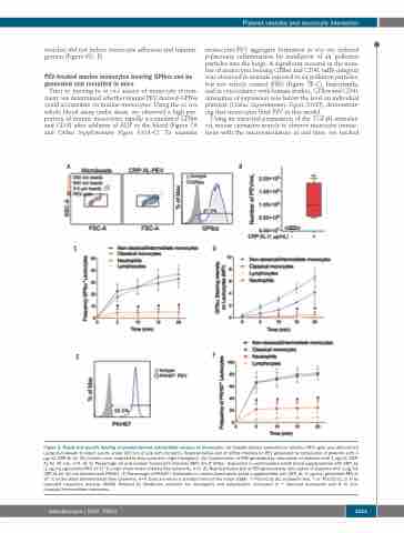
Figure 3. Rapid and specific binding of platelet-derived extracellular vesicles to monocytes. (A) Platelet-derived extracellular vesicles (PEV) gate was determined using microbeads to select events under 900 nm in size (left dot plots). Representative plot of GPIbα intensity on PEV generated by stimulation of platelets with 1 μg/mL CRP-XL for 30 minutes (min) analysed by flow cytometry (right histogram). (B) Concentration of PEV generated by stimulation of platelets with 1 mg/mL CRP- XL for 30 min, n=4. (C, D) Percentage (C) and median fluorescent intensity (MFI) (D) of GPIbα+ leukocytes in unstimulated whole blood supplemented with CRP_XL (1 mg/mL)-generated-PEV at 37°C under shear determined by flow cytometry, n=4. (E) Representative plot of PEV generated by stimulation of platelets with 1 mg/mL CRP-XL for 30 min labelled with PKH67. (F) Percentage of PKH67+ leukocytes in unstimulated whole blood supplemented with CRP_XL (1 mg/mL) generated PEV at 37°C under shear determined by flow cytometry, n=4. Data are mean ± standard error of the mean (SEM). **P≤0.01 by (B) unpaired t-test. * or #P≤0.01 (C, D, F) by repeated measures two-way ANOVA followed by Bonferroni post-test for neutrophils and lymphocytes compared to * classical monocytes and # to non- classical/Intermediate monocytes.
haematologica | 2020; 105(5)
1253
