
Page 92 - Haematologica May 2020
P. 92
M. Chimen et al.
tion contributed to GPIbα accumulation. For these stud- ies, we used CRP-XL to stimulate whole blood, as this agonist does not directly activate monocytes and would thus allow analysis of whether secondary activation of monocytes downstream of platelet activation was prereq- uisite for PEV adhesion. We assessed the expression of the activation marker αMβ2-integrin (CD11b/CD18) on monocytes 30 min after the addition of CRP-XL to whole blood. There was some increase in both integrin subunits CD11b and CD18 (Online Supplementary Figure S9C-D), however, this was inconsistent and monocyte subset spe- cific. When a function neutralising antibody against CD18 was included in the assay it had no effect on GPIbα accumulation (Figure 4C-E), indicating that monocyte activation was not required for PEV adhesion.
Adopted GPIbα is a functional adhesion molecule supporting monocyte rolling on von Willebrand Factor
cytes was inhibited by a function-neutralising antibody against GPIbα (Figure 5D, E).
As GPIbα is known to mediate binding of platelets from flowing blood to von Willebrand Factor (VWF), we tested whether VWF could also recruit PEV-treated mono- cytes (Figure 5A-E). Monocytes lacking GPIbα showed low levels of adhesion when perfused across immobilised human VWF (Figure 5B, E). However, acquisition of PEV- derived-GPIbα supported capture and rolling (66.8±4.1% of adherent cells rolling) of monocytes on VWF (Figure 5C, E). Importantly, the adhesion of PEV-treated mono-
Transforming growth factor beta-1 (TGF-β1) promotes the expression of a matrix of VWF on the surface of EC which recruits platelets from flowing blood, which in turn function as adhesive bridges for the preferential recruitment of monocytes to EC in vitro and in vivo.15 Here we used this model to determine whether PEV-derived GPIbα could support monocyte adhesion directly to stimulated endothelium. A low level of monocyte adhe- sion to TGF-β1-stimulated EC was observed without PEV (Figure 6A, D). However, PEV-treated monocytes adhered in significantly higher numbers, an adhesive interaction blocked by a GPIbα blocking antibody (Figure 6B-D). As previously observed, recruited mono- cytes did not roll on the EC. Thus 6.1±0.9% of adherent cells were observed rolling, with the remaining 93.9% becoming activated and stably adherent. Interestingly, the acquisition of PEV increased the efficiency with which monocytes transmigrated across the EC monolay- er (Figure 6E). We could attribute this increase in PEV- treated monocytes recruitment to PEV rather than solu- ble factors such as chemokines, as supernatants generat- ed from PEV filtered using a 10 KDa size filter (to remove
A
B
CD
Monocytes bearing GPIbα bind to EC in a model of vascular inflammation
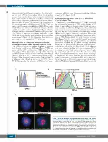
Figure 2. GPIbα on monocytes in stimulated whole blood derives from platelet- derived extracellular vesicles. (A, B) Representative plots of GPIbα labelling on monocytes (all) in unstimulated or TRAP (100 mM)-stimulated whole blood under shear for 30 minutes (min) (A) and over time (B) measured by flow cytometry. (C) Representative pictures of monocytes labelled with anti-CD14, anti-GPIbα for platelet-derived extracellular vesicles (PEV) and DAPI imaged by confocal microscopy. Monocytes were isolated and incubated with CRP-XL-generated PEV for 30 min at 37°C under shear prior fixing and labelling. (D) Monocytes bound to platelets, both labelled with FITC-phalloidin and DAPI imaged by confocal microscopy.
1252
haematologica | 2020; 105(5)
