
Page 94 - Haematologica May 2020
P. 94
M. Chimen et al.
human mouse PEV-treated monocytes (Online Supplementary Figure S10E-F). We used the hIL4R/GPIbα−Tg mouse which expressed human IL-4 receptor under the GPIbα promoter. This allows the ani- mals to be rendered thrombocytopenic by injection of an antibody against hIL4R. Adoptively transferred WT platelets or PEV are however retained within the circula- tion. Using mice depleted of endogenous platelets using an anti-hIL4R antibody, we observed higher numbers of adoptively transferred WT PEV-treated monocytes rolling on the microvasculature compared to untreated mono- cytes; the number was significantly reduced by a GPIbα blocking antibody (Figure 7D-G). Detailed analysis revealed two populations of rolling cells: those exhibiting stable rolling (interactions >300 ms) with a velocity of 241±82 mm/s (Figure 7D, F); those exhibiting transient rolling (interactions <300 ms) with a velocity of 478±65 mm/s (Figure 7D, G). We also infused human monocytes
AB
into ApoE-/- mice that had been on a western diet for six weeks and observed the carotid artery by intravital microscopy. Murine PEV-treated monocytes adhered to the artery wall with significantly greater efficiency than untreated monocytes (Figure 7H). In this environment a mixture of adhesive behaviors was observed with station- ary adhesion, stable rolling and transient rolling adhesion evident (Figure 7H).
Monocytes with platelet markers appear within 1 hour of severe trauma and are rapidly cleared from the circulation
We investigated whether rapid production and binding of extracellular vesicles to monocytes could be detected following an acute event such as traumatic injury. In the Golden Hour study blood samples in the pre-hospital set- ting (mean time to blood sampling =43 min) were acquired from traumatically-injured patients (injury sever-
CDE
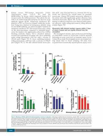
Figure 4. Blocking of GPIbα+ platelet-derived extracellular vesicles to leukocytes. (A) Binding of platelet-derived extracellular vesicles (PEV) on classical monocytes, non-classical/intermediate monocytes and neutrophils with blockade of P-selectin in TRAP (100 mM)-stimulated whole blood for 30 minutes (min) under shear, n=3. (B) Surface expression (MFI: median fluorescence intensity) of PSGL1 (P-selectin ligand) on monocyte subsets, neutrophils and lymphocytes determined by flow cytometry, n=3. (C-E) Binding of PEV on classical monocytes (C), non-classical/intermediate monocytes (D) and neutrophils (E) with blockade of CD31, ICAM-2, CD18 (β2) and Phosphatidylserine (PS) in TRAP (100 mM)-stimulated whole blood for 30 min under shear, n=3-5. Data are mean ± standard error of the mean (SEM). *P≤0.05, **P≤0.01 compared the normalised IgG control (A) by analysis of variance (ANOVA) and Dunnett post-test or Bonferroni post-test (B) and one sample t-test to 100% of TRAP control (C-E).
1254
haematologica | 2020; 105(5)
