
Page 153 - Haematologica May 2020
P. 153
TARP as target in acute myeloid leukemia
T-cell receptor γ chain alternate reading frame protein is expressed in acute myeloid leukemia cell lines and patient leukemic cells
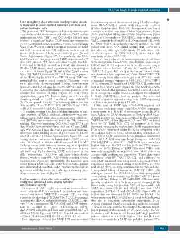
We generated TARP transgenic cell lines in order to opti- mize western blot experiments and evaluate TARP protein expression in AML. THP-1 and OCI-AML3 OE cell lines showed a significant higher TARP transcript expression (P<0.01) compared to mock controls (Online Supplementary Figure S6A). Western blotting confirmed presence of TARP and GFP proteins in both OE cell lines, with a size of around 20 kDa and 27 kDa, respectively (Figure 2A and Online Supplementary Figure S7). Concordantly, the OCI- AML3 mock cell line, negative for TARP, only showed a 27 kDa GFP protein. WT AML cell lines HL-60, MV4;11, THP-1 and MOLM-13, as well as LNCaP, also showed a 20 kDa TARP protein, with expression corresponding to the transcript levels (Figure 2B and Online Supplementary Figure S7). TARP knockdown (KD) cell lines were generat- ed for HL-60, Kg-1a, MV4;11 and THP-1 using TARP-tar- geting shRNA, next to mock controls. Transcript levels were efficiently down-regulated (Online Supplementary Figure S8), and KD cell lines for HL-60, MV4;11 and THP- 1 showing the highest transcript downregulation were selected for western blotting (Figure 2C and Online Supplementary Figure S7). Protein levels were efficiently down-regulated in HL-60 transduced with shRNA 3 (19.4% compared to mock). This downregulation was less clear in MV4;11 and THP-1: 116% (shRNA 3) and 108% (shRNA 3) versus 63% (shRNA 2), respectively.
To confirm western blot data and determine the subcel- lular location of TARP, confocal microscopy was per- formed using TARP antibodies combined with mitochon- drial (HSP-60) and endoplasmic reticulum (ER, calnexin) staining. The over-expressing OCI-AML3 and THP-1 cell lines (Online Supplementary Figure S6B and C) and TARP- high WT AML cell lines showed a perinuclear membra- nous-type TARP staining pattern (Kg-1a (Figure 3), HL-60, MV4;11 and THP-1 (Online Supplementary Figure S9). This finding was in contrast to the barrel-shaped TARP pattern with mitochondrial co-localization reported in LNCaP.43 Co-localization with calnexin, presenting as a speckled pattern throughout the ER, was more abundant in some cell lines, e.g. Kg-1a, showing TARP enrichment at the cells’ protrusions. TARP-low cell lines concordantly showed weak or negative TARP protein staining (Online Supplementary Figure S8). Importantly, the leukemic cells sorted from a TARP-high and TARP-low pedAML patient also illustrated differential TARP protein expression in agreement with the transcript levels, again showing limit- ed mitochondrial overlap (Figure 3).
T-cell receptor γ chain alternate reading frame protein transgenic cytotoxic T cells display specific anti-leukemic activity
To explore if TARP might represent an immunothera- peutic target in AML, we evaluated the cytokine and cyto- toxicity response of TARP-TCR transgenic CTL, encoding a previously developed TCRA8-T2A-TCRB12 sequence targeting the HLA-A2 enhanced affinity TARP(P5L)4-13 epi- tope.46,47 As concomitant HLA-A*0201 and TARP expres- sion is required to trigger TCR-mediated killing, HLA-A*0201 transgenic cell lines were generated for 3 WT cell lines (HL-60, Kg-1a and MOLM-13) and 3 Luc-positive cell lines (HL-60-Luc, MOLM-13-Luc, MV4;11-Luc).
First, target specificity of the TARP-TCR was examined
in a non-competitive environment using T2 cells (endoge- nous HLA-A*0201+) pulsed with exogenous peptides (Online Supplementary Table S3). As expected, we found stronger cytokine responses (Online Supplementary Figure S10A) and higher killing rates (Online Supplementary Figure S10B and C) towards the TARP(P5L)4-13 than to the cognate TARP4-13 peptide for both RV and LV transduced CTL, with LV TARP-TCR CTL generally reacting stronger. T2 cells pulsed with non-TARP-related peptides (INF, CMV) were not affected, although CMV-pulsed T2 cells were effi- ciently recognized by CMV-TCR CTL, indicating a high specificity of the TARP-TCR.
Second, we explored the immunogenicity of cell lines with endogenous HLA-A*0201 presentation. Exposure to LNCaP and THP-1 appeared to be insufficient to trigger cytokine release for both LV and RV transduced TARP- TCR CTL (Figure 4A). Using a chromium51 release assay, we observed a lytic response by LV transduced TARP-TCR CTL starting from effector to target ratio (E/T) 10/1, with a maximal average response at 50/1 (LNCaP 10%, THP-1 24%), whereas RV transduced TARP-TCR CTL performed best at 10/1 (THP-1 12%) (Figure 4B). The TARP-low AML cell line OCI-AML3 remained unaffected under all condi- tions. Altogether, as the TARP-TCR targets the enhanced HLA-A2 TARP(P5L)4-13 binding peptide, we observed weaker responses against endogenous TARP-expressing cell lines compared to pulsed T2 cells.
Third, lysis of TARP-high HLA-A*0201-negative cell lines was evaluated versus their HLA-A*0201 transgenic counterparts in a 48-h FCM-cytotoxicity assay. In addi- tion, killing of TARP transgenic or TARP-pulsed HLA- A*0201-positive cell lines was compared to the respective TARP-low WT cell line (Figure 4C). A non-TARP mediated lysis by LV TARP-TCR CTL of maximal 20% was observed (indicated by dashed line). Stable transduction of HLA-A*0201 increased killing for Kg-1a compared to the WT cell line (29% vs. 13%), whereas killing of MOLM-13, with lower TARP expression levels, remained unaffected when HLA-A*0201 was introduced. Transgenic TARP OE and TARP(P5L)4-13 pulsed OCI-AML3 cells were prone to a higher lysis than the WT cell line (44% and 55%, respec- tively, vs. 24%). Killing of TARP OE/pulsed THP-1 cells was only marginally up-regulated, most likely due to an already high endogenous expression. These data were confirmed using RV TARP-TCR CTL, and corrected for non-TARP mediated lysis using mock CTL. HLA-A*0201 expression again increased killing of Kg-1a (46% vs. -4%) and HL-60 (40% vs. 15%) compared to the WT cell line. Up-regulated killing of transgenic TARP OE THP-1 cells was again limited. For OCI-AML3, lysis was up-regulated after pulsing, but remained low for the TARP OE trans- genic cell line. Killing by LV TARP-TCR CTL was addi- tionally evaluated in a bioluminescence imaging (BLI)- based assay using Luc-positive AML cell lines with high TARP expression (HL-60 and MV4;11) and low TARP expression (MOLM-13 and OCI-AML3) (Figure 4D). A higher lysis was observed for HL-60-Luc and MV4;11-Luc when expressing HLA-A*0201 at 48 h and 56 h, indicating that also in long-term cytotoxicity experiments HLA- A*0201 restricted TARP-specific killing could be detected.
Finally, we explored the feasibility of therapeutic target- ing of primary leukemic cells by LV TARP-TCR CTL. Co- incubation with blasts sorted from a TARP-high pedAML patient resulted into a 2-fold higher IFN-γ and IL-2 pro- duction compared to a TARP-low pedAML patient (22%
haematologica | 2020; 105(5)
1313
