
Page 151 - Haematologica May 2020
P. 151
TARP as target in acute myeloid leukemia
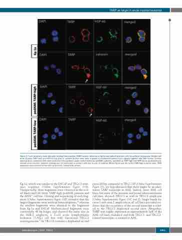
Figure 3. T-cell receptor γ chain alternate reading frame protein (TARP) protein detection in Kg-1a and patient leukemic cells by confocal microscopy. Merged pat- terns visualize TARP (red) and HSP-60 (top lane) or calnexin (bottom lane) (both in green) co-localization (yellow fusion signals) together with DAPI nuclear counter- staining (blue). Leukemic cells were sorted from two pediatric acute myeloid leukemia (pedAML) patients, classified as TARP-high and TARP-low by qualitative poly- merase chain reaction. Calnexin staining was not performed on primary cells due to lack of material. Within Kg-1a and the sorted TARP-high leukemic cells, TARP expression was enriched at the cells’ protrusions, indicated by arrows.
Kg-1a, which was similar to the LNCaP and TRGC1 refer- ence sequence (Online Supplementary Figure S5A). Unexpectedly, three fragments were observed in the sort- ed blasts and LSC from TARP-high pedAML patients and the MV4;11 cell line. Cloning and sequencing of each frag- ment (Online Supplementary Figure S5B) revealed that the largest fragments were artificial heteroduplexes,44 whereas the smallest fragments were identical to the fragments from Kg-1a and LNCaP. Medium-sized fragments were consistently 48 bp longer, and showed the same size as the HSB-2 amplicon, a T-cell acute lymphoblastic leukemia (T-ALL) cell line with functional TRGC2 rearrangements.45 As TRGC2 contains a duplicated second
exon (48 bp) compared to TRGC145 (Online Supplementary Figure S2), we hypothesized that there might be an alter- native TARP transcript in AML. Indeed, most AML cell lines, but none of the prostate and breast adenocarcinoma cell lines, showed TRGC1 as well as TRGC2 amplicons (Online Supplementary Figure S5C and E). Single bands for exon 3 and exon 1 amplicons in all cell lines provided evi- dence that the occurrence of the second transcript is relat- ed to the TRGC2 duplicated second exon. Altogether, TARP was highly expressed in approximately half of the AML cell lines evaluated, and both TRGC1- and TRGC2- related transcripts co-existed in AML.
haematologica | 2020; 105(5)
1311
