
Page 152 - Haematologica May 2020
P. 152
B. Depreter et al.
AB
C
DE
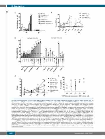
Figure 4. Functional evaluation of T-cell receptor (TCR)-transgenic cytotoxic T cells (CTL) towards cognate and modified cell lines and patient leukemic cells. (A) Cytokine response (IFN-γ/IL-2 expression within the CD3+/CD8+ compartment) by co-incubation (1 hour, h) with OCI-AML3 and THP-1 was evaluated by both lentiviral (LV) and retroviral (RV) TCR-T-cell receptor γ chain alternate reading frame protein (TARP) CTL. LNCaP and patient leukemic cells (single experiment) were only eval- uated by LV transduced TARP-TCR CTL. For each target, positive (+) or negative (-) HLA-A*0201 and TARP expression, in this respective order, is indicated between brackets. HLA-A*0201 and TARP co-expressing cell lines (LNCaP and THP-1) were unable to trigger higher cytokine release than OCI-AML3 with low TARP expression. Leukemic cells from a TARP-high pediatric acute myeloid leukemia (pedAML) patient triggered a 2-fold higher cytokine release compared to a TARP-low pedAML patient. (B) Lytic response of LV and RV TARP-TCR CTL versus HLA-A*0201-positive TARP-high (black symbols) and TARP-low (white symbols) targets, measured by a chromium51 release assay after 4 h. Highest lysis of TARP-high cell lines was observed at E/T ratio 50/1 for LV and 10/1 for RV TARP-TCR CTL (percentages indicated between brackets), whereas OCI-AML3 (HLA-A*0201+, TARP–) remained unaffected. (C) Lytic response of LV and RV TARP-TCR CTL versus towards wild-type (WT), transgenic and pulsed AML cell lines, measured by a 48-h FCM-based cytotoxicity assay. The dashed line indicates the highest level of non-TARP mediated back- ground killing observed for LV TARP-TCR CTL, as no mock CTL could be constructed. Positive (+) or negative (-) expression for HLA-A*0201 and TARP is shown, in this respective order, between brackets. Bold symbols indicate the expression differing from wild-type, either by retroviral transduction or pulsing. HLA-A*0201 transgenic AML cell lines were more efficiently lysed compared to their HLA-A*0201-negative counterparts (Kg-1a, MOLM-13, HL-60). Higher lysis was observed for transgenic TARP OE or peptide-pulsed cell lines compared to the WT cell line (OCI-AML3, THP-1), except for killing of TARP OE OCI-AML3 cell line by RV TARP-TCR CTL. (D) Lysis by LV TARP-TCR CTL, measured at different time points (8h, 24h, 48h and 56h, as indicated on x-axis), based on the luminescence release by transgenic HLA-A*0201- expressing TARP-high AML cell lines with respect to the HLA-A*0201 WT cell line (HL-60-Luc, MOLM-13-Luc and MV4;11-Luc: black symbols). In addition, lysis of the TARP-low, cognate HLA-A*0201-positive OCI-AML3 cell line was evaluated (white symbols). Mean lysis (%) observed after 48 h is indicated next to whiskers, repre- senting the mean±standard error of mean. (E) 48-h FCM-based cytotoxicity assay evaluating lysis of primary leukemic cells (adult AML=5, all FLT3-ITD mutated) by LV TARP-TCR transduced CTL (biological duplicates). TARP transcript expression (CNRQ) is shown in the x-axis for each target. IFN-γ: interferon gamma; IL-2: inter- leukin-2; INF: influenza.
1312
haematologica | 2020; 105(5)
