Page 260 - Haematologica April 2020
P. 260
S.I. Obydennyi et al.
μM) significantly diminished the PS+ fraction formed by the WAS platelets on fibrinogen (Figure 4A). Other inhibitors, including necroptosis inhibitor necrostatin-1, calpain inhibitor calpeptin and pan-caspase inhibitor Z- VAD-FMK, had no significant effect on PS exposure (Figure 4B). These data strongly suggest that it is indeed the necrotic mechanism of mitochondrial permeability transi- tion pore opening that is responsible for increased PS expo- sure in WAS platelets.
Role of calcium homeostasis, cellular energetics, and reactive oxygen species in phosphatidylserine exposure by Wiskott-Aldrich syndrome platelets
In order to get further insight into the necrosis of the sur- face-attached WAS platelets, they were treated with xestospongin C (an inositol trisphosphate receptor block- er), or thapsigargin (a sarco-endoplasmic reticulum Ca2+ ATPase inhibitor), or in buffer A without addition of calci- um chloride (Figure 4C). Xestospongin C inhibited PS exposure suggesting involvement of inositol trisphosphate signaling, while thapsigargin potently boosted platelet necrosis. In contrast, the effects were drastically decreased in the absence of extracellular calcium.
Importantly, thapsigargin caused accelerated cell death in the WAS platelets compared with platelets from healthy controls in suspension as well without any surface attach- ment (Figure 4D), which suggests that the WAS platelets’ propensity to necrosis is caused by dysregulation of their calcium homeostasis. The same experiment with lactad- herin and without addition of extracellular calcium did not show an increased PS+ fraction of WAS platelets (Figure 4E). For an additional check of the effect of outside-in sig- naling on thapsigargin-induced PS exposure in this design, we pre-treated platelets with the integrin αIIbβ3 antagonist monafram which did not affect the thapsigargin-induced PS exposure (Online Supplementary Figure S4). Pre-incuba- tion of the WAS platelets with the mitochondrial ATPase inhibitor oligomycin or with the mitochondrial uncoupler CCCP increased the formation of PS+ platelets at thapsigar- gin treatment in the case of WAS platelets, while the mito- chondrial respiratory chain complex I inhibitor rotenone had less effect on the thapsigargin-induced PS exposure (Figure 4F); none of these three drugs caused platelet necro- sis by themselves. These data indicate that an energy defi- ciency could be a factor contributing to platelet necrosis but not the defining one. In line with this, although the lev- els of ATP in cells were decreased in parallel with the increase of the PS+ platelets upon thapsigargin treatment, the same decrease of ATP was caused by CCCP without PS exposure indicating that the observed phenomenon is not purely caused by an energy collapse (Figure 4G, H). ROS production in the WAS platelets was not essentially different from that in healthy donor platelets, and was only mildly increased upon stimulation with CRP (Online Supplementary Figure S5). The morphology of the mito- chondria in WAS platelets was not apparently different from that of normal ones, as judged by transmission elec- tron microscopy (Online Supplementary Figure S6).
Platelet necrosis correlates directly with the number of mitochondria
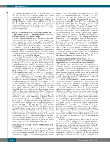
During examination of the images, it became apparent that the WAS platelets undergoing PS exposure and mito- chondrial membrane potential loss rarely had more than two mitochondria per cell. We, therefore, performed exper-
iments to count the number of mitochondria in each platelet and correlated this with the outcome (i.e. PS expo- sure) (Figure 5). For both WAS patients and healthy donors, the number of mitochondria was significantly lower in the platelets that became PS+ (Figure 5A). This number affected the fate of platelets in a dose-dependent manner: about 33% of the WAS platelets exposed PS if they had one to four mitochondria per platelet, and only about 11% if they had more than five mitochondria (Figure 5B). A similar dependence was observed for platelets from healthy donors (Figure 5B), although they exposed PS more rarely. The his- togram in Figure 5C shows the distributions of mitochon- dria number for platelets from WAS patients and healthy donors side by side. Importantly, although the mean num- ber of mitochondria in WAS platelets was not much lower than that in the control platelets, there was significant skewing to the left of the curve: a total of 27±12% of WAS platelets had fewer than three mitochondria, compared to only 8.7±4.4% of healthy platelets. In order to check if the number of mitochondria has a wider significance in platelet necrosis, we performed experiments with fibrinogen- attached healthy platelets stimulated with TRAP-6 or thrombin, revealing the same pattern (Figure 5D, E).
Systems biology simulations reveal critical roles of mitochondrial number and surface-to-volume ratio in programmed cell death in Wiskott-Aldrich syndrome
In order to dissect the mechanisms of mitochondria- dependent necrosis in WAS, we developed a computation- al systems biology model of calcium signaling (Figure 6). In the model, which incorporated all compartments and major calcium signaling mechanisms, we investigated dependence of the platelet calcium response on two major variables that differ for the WAS platelets, the number of mitochondria and platelet size.
The model demonstrated that a decrease in the number of mitochondria should make platelets more sensitive to mitochondrial collapse and result in a higher increase in calcium because the remaining mitochondria could not bear the ATP production load (Figure 6A, B), which agrees well with the experimental observations. We also simulat- ed platelets of different sizes; when scaling them, the ratio between surface and volume molecules was naturally changed (Figure 6), as volume is proportional to the size to the third degree, while surface is proportional to the size to the second degree. Upon stimulation, the virtual platelets with a smaller size had comparable active phospholipase C per volume (Figure 6C), but more inositol trisphosphate and ultimately much more calcium (Figure 6E) because they had more inositol trisphosphate receptors per volume (as these were assumed to be proportional to the surface). This is again in line with the experimental data presented above that showed increased calcium levels in WAS even prior to mitochondrial permeability transition pore open- ing, and with the sensitivity of the phenomenon to xestospongin C.
The model, therefore, predicted that the size of platelets from untreated WAS patients would negatively affect the platelets’ ability to expose PS spontaneously and (if this is the mechanism underlying thrombocytopenia) positively affect the patients’ platelet count. Interestingly, there was a significant positive correlation between platelet size and platelet count among the untreated WAS patients (Online Supplementary Figure S7A). Although we did not observe significant correlations with PS exposure, probably as a
1102
haematologica | 2020; 105(4)


