
Page 258 - Haematologica April 2020
P. 258
S.I. Obydennyi et al.
(Online Supplementary Figure S3A, time point 116 s). If it was reversed, calcium concentration also decreased (Online Supplementary Figure S3A, time point 146 s). This is drastically different from healthy donors' activated platelets, in which calcium was not so sensitive to the col- lapse of a single mitochondrion.18 Interestingly, WAS platelets with large numbers of mitochondria had increased background cytosolic calcium and frequent oscillations, but were not sensitive to the collapse of sin- gle mitochondria either (Online Supplementary Figure S3B).
Phosphatidylserine exposure on Wiskott-Aldrich syndrome platelets is mediated by mitochondrial permeability transition pore opening
The critical element of mitochondrially driven necrosis is mitochondrial permeability transition pore opening. To check this, we added several inhibitors of different cell death-regulating signaling pathways during platelet incu- bation on fibrinogen (cyclosporine A, necrostatin-1, Z- VAD-FMK) or spreading (calpeptin). The mitochondrial permeability transition pore inhibitor cyclosporine A (5
AB
CD
E
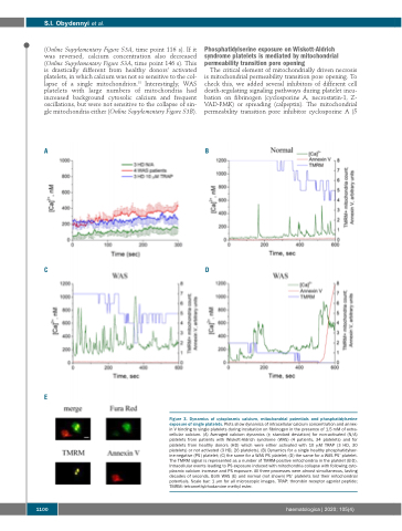
Figure 3. Dynamics of cytoplasmic calcium, mitochondrial potentials and phosphatidylserine exposure of single platelets. Plots show dynamics of intracellular calcium concentration and annex- in V binding to single platelets during incubation on fibrinogen in the presence of 1.5 mM of extra- cellular calcium. (A) Averaged calcium dynamics (± standard deviation) for non-activated (N/A) platelets from patients with Wiskott-Aldrich syndrome (WAS) (4 patients, 34 platelets) and for platelets from healthy donors (HD) which were either activated with 10 μM TRAP (3 HD, 30 platelets) or not activated (3 HD, 26 platelets). (B) Dynamics for a single healthy phosphatidylser- ine-negative (PS-) platelet; (C) the same for a WAS PS- platelet; (D) the same for a WAS PS+ platelet. The TMRM signal is represented as a number of TMRM-positive mitochondria in the platelet (B-D). Intracellular events leading to PS exposure induced with mitochondria collapse with following cyto- plasmic calcium increase and PS exposure. All three processes were almost simultaneous, lasting decades of seconds. Both WAS (E) and normal (not shown) PS+ platelets lost their mitochondrial potentials. Scale bar: 1 μm for all microscopic images. TRAP: thrombin receptor agonist peptide; TMRM: tetramethylrhodamine methyl ester.
1100
haematologica | 2020; 105(4)
