
Page 256 - Haematologica April 2020
P. 256
S.I. Obydennyi et al.
PS+ platelets under resting conditions and normal PS expression upon stimulation (Online Supplementary Figure S2). In summary, WAS platelets were small and demon- strated some decreased preactivation features in the rest- ing state but they did not appear to have any drastic func- tional differences from normal platelets in an activated state.
Signaling events in single fibrinogen-attached Wiskott-Aldrich syndrome platelets
To identify the mechanisms of the PS externalization of the WAS platelets, we simultaneously examined dynam- ics of calcium in the cytosol, mitochondrial membrane potential and PS exposure in WAS and control platelets (Figure 3). The mean intracellular cytosolic calcium level in the WAS platelets was 4-fold greater than that in the control platelets at the beginning, and the average differ-
ence increased with time (Figure 3A). While healthy
unstimulated platelets, in line with previous reports,18,32
had only occasional calcium spikes when bound to fib-
rinogen (Figure 3B), unstimulated platelets from WAS
patients had frequent oscillations with longer spike dura-
tion (Figure 3C, D). The mitochondria in the WAS
platelets lost their membrane potential one after another
and, if all of them became TMRM-negative, the cell
began to bind annexin V within 10 s (Figure 3D), exactly
as reported before for the PS+ platelet formation induced
in healthy donors with TRAP-6 or thrombin.18,27 This is in
agreement with the scenario of mitochondrial calcium-
overloading-induced necrosis of procoagulant platelet for- mation.18,33,34
Importantly, time lapse imaging revealed that mito- chondrial collapse in WAS platelets with few mitochon- dria in turn led to a rapid cytosolic calcium increase
AB
C
D
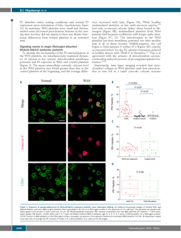
Figure 1. Exposure of phosphatidylserine by Wiskott-Aldrich syndrome platelets upon fibrinogen binding. (A) Confocal microscopy images of healthy (left) and Wiskott-Aldrich syndrome (WAS) (right) platelets after spreading for 30 min on a fibrinogen surface in the presence of 1.5 mM Ca2+. The platelets are labeled with CD61 (green) and annexin V (red); scale bar: 10 μm. (B) Phosphatidylserine-positive (PS+) fraction of platelets from the WAS patients (27 patients, >7,500 cells), adult healthy (18 donors, >6,500 cells) and 0- to 7-year old children without WAS (6 children, age: 0, 0, 2, 3, 4, 7 years, 2,300 platelets) on a fibrinogen surface. (C) PS+ fraction of WAS platelets on the fibrinogen surface, showing a comparison of romiplostim-treated and untreated WAS patients, P=0.94. (D) Monafram-coated coverslips did not change the PS+ fraction, P=0.86, n=4, 2,500 platelets. w/o: without; FG: fibrinogen.
1098
haematologica | 2020; 105(4)
