
Page 259 - Haematologica April 2020
P. 259
Programmed necrosis of platelets in WAS
AB
CD
EF
GH
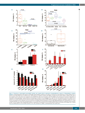
Figure 4. Prevention of mitochondrial permeability transition pore opening affects spontaneous phosphatidylserine exposure of Wiskott-Aldrich syndrome platelets. (A) Phosphatidylserine (PS) exposure of fibrinogen-spread platelets incubated in the absence or presence of 5 μM cyclosporine A (3,900 platelets from 11 patients and 4,000 platelets from 6 healthy donors). (B) PS exposure of platelets incubated with DMSO or programmed cell death inhibitors calpeptin (200 μM, 20 min incubation), Nec-1 (50 μM, 50 min incubation) and Z-VAD-FMK (50 μM, 50 min incubation) (100-300 platelets were observed for each dot). (C) Modulation of intracellular calcium signaling in spread platelets by xestospongin C (3 μM, 50 min); thapsigargin (TG, 1 μM, 30 min); with lactadherin and without addition of 1.5 mM CaCl2 (n=5, 6,800 platelets). (D) Flow cytometry analysis of Wiskott-Aldrich syndrome (WAS) platelet PS exposure. Incubation of platelets in suspension with 1 μM TG in the presence of 1.5 mM CaCl2 for 10 min induced a PS+ platelet fraction comparable to that with fibrinogen-spreading for both WAS patients (n=7, mean ± standard deviation: 19.7%±11.8%) and healthy donors (n=11, 6.6%±8.0%). (E) Flow cytometry analysis of WAS platelet PS exposure in suspension without addition of CaCl2 (n=3); (F) Analysis of the mitochondrial inhibitors in suspension at TG treatment (n=3); (G, H) ATP levels (G) versus the number of PS+ platelets (H): in healthy donors and WAS patients at TG and CCCP treatment (n=3). CsA: cyclosporine A, DMSO: dimethylsulfoxide; HD: healthy donor.
haematologica | 2020; 105(4)
1101
