
Page 247 - Haematologica March 2020
P. 247
SOX11 and TP53 status in MCL
caveat of any RNA-based technology including RNAscope is that good mRNA preservation is needed, and this depends on good fixation procedures. However, the low dropout rate in our study, with only two decalcified BM specimens not evaluable based on the positive control, documents the robust performance of RNAscope in rou- tine archival tissue specimens.
An intriguing finding in this study was the higher fre- quency of TP53 mutations in cases with low/negative SOX11 mRNA levels both in cMCL as well as nnMCL. TP53 mutations are rarely observed in MCL with classic morphology,11 but are frequently found in highly prolifer- ative MCL with blastoid morphology (up to 30%), and associated with aggressive clinical course.29,30,49 A recent study comparing the gene signature of cMCL and nnMCL demonstrated that the incidence of TP53 mutations is sim- ilar in both subgroups (36% and 38%, respectively).50 Nevertheless, an interesting observation was the report of a group of cMCL cases that lacks SOX11 expression, car- ries TP53 mutations and has a dismal prognosis.11 This finding was recently confirmed in a study from the European MCL network that demonstrated that p53 over- expression is preferentially found in SOX11 negative cMCL cases (50% vs. 13%).26 A caveat of the latter study
is that molecular analysis to demonstrate the presence of TP53 mutations was not performed. Although, p53 stain is a very good surrogate marker of gene mutation when strong, homogeneous staining is considered as positive, a group of cases with negative or low p53 staining due to deletions or frameshift mutations are missed as demon- strated in this study (2/13; 15%). Accordingly, of the TP53 mutated cases nine were in the negative/low SOX11 mRNA level group and four in the high mRNA level group (60% vs. 11%, P=0.0007). Our results further confirm pre- vious reports suggesting that TP53 overexpression/muta- tions frequently occur in SOX11 negative/low cases.11,26 It is worth mentioning that in the two previous studies only nodal cMCL cases with no previous history of leukemic manifestation were included. Nevertheless, it has been suggested that these cases might correspond to a selected subset of progressed nnMCL tumors.17,48 However, con- trary to the cases reported by Nygren et al. and Aukema et al., in this study eight nnMCL cases with typical presenta- tion diagnosed in the BM and one in spleen were included. Although the TP53 mutated nnMCL cases showed strik- ing lymphocytosis in the peripheral blood, they lacked sig- nificant lymphadenopathy and rarely blastoid morpholo- gy was observed. The high incidence of nnMCL with
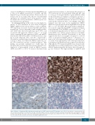
AB
CD
Figure 4. Mantle cell lymphoma (MCL) with blastoid morphology, low SOX11 expression and TP53 mutation (case # 58). (A) the infiltrate is characterized by a dif- fuse, monotonous population of medium-sized to large cells with irregular nuclei, fine chromatin and narrow cytoplasm (hematoxylin and eosin); (B) p53 is strongly positive in the majority of the tumor cells; (C) the SOX11 stain shows a rather weak heterogeneous positivity in the tumor cells (<10%); (D) SOX11 RNAscope shows a score 1 with 1-3 dots/cell and no dot clusters. All original magnification 200x.
haematologica | 2020; 105(3)
761
