
Page 243 - Haematologica March 2020
P. 243
SOX11 and TP53 status in MCL
Correlation between SOX11 and TP53 status
Table 3 and Online Supplementary Table S1 summarize the data of the TP53 mutated group. In thirty-seven cases, all exons of TP53 were sequenced by NGS including all p53 positive cases (11 cases), the five equivocal cases and 20 p53 negative cases including all remaining blastoid cases. TP53 mutations were demonstrated in 13 cases; 11 considered p53 positive, and two considered p53 nega- tive; one of these a nnMCL case revealed complete nega- tivity by IHC. The five equivocal and all remaining nega- tive cases were TP53 wild-type. Although the cut-off for
p53 positivity was set on >20%, 10 cases showed strong p53 expression in more than 60% of the tumor cells. TP53 mutations were identified in eight of 56 cMCL cases (14%), five of them with blastoid morphology (Figure 4). Four of these eight cases were in the group of negative/low SOX11 expression. In contrast, five of seven evaluable cases (71%) of nnMCL had TP53 mutations (14% vs. 71%; P=0.003). Four of these cases were diag- nosed in BM and presented with striking lymphocytosis in the peripheral blood (Figure 5). One case had a blastoid morphology. Altogether, nine of the TP53 mutated cases
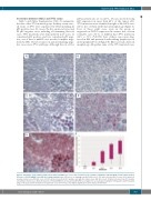
AB
CD
E
F
Figure 1. Illustration of the staining pattern in the different RNAscope scores and correlation to the real-time quantitative PCR (RT-qPCR) results, which shows a decrease of SOX11-mRNA levels with decreasing staining scores. (A) score 0, no staining (case #16); (B) score 1, 1-3 dots/cell (case #9); (C) score 2, 4-9 dots/cell, very few clusters (case #5); (D) score 3, 10-15 dots/cell, less than 10% positive cells with dot clusters (case #2); (E) score 4, 15 dots/cell, more than 10% positive cells with dot clusters (case #11); (F) box plot of SOX11 mRNA levels measured by RT-qPCR according to the score groups showing the third quartile and first quartile range of the data and the median for the particular score (P=0.0002). A-E original magnification 200x and insert with 400x.
haematologica | 2020; 105(3)
757
