
Page 234 - Haematologica March 2020
P. 234
A. Falgàs et al.
Firstly, we evaluated CXCR4 expression in the mouse B-cell lymphoma WEHI-231 cell line that showed medium CXCR4 membrane expression by flow cytometry and IHC (Online Supplementary Figure S2A). Then, we demon- strated intracellular nanocarrier uptake in mouse WEHI- 231 cells and its dependence on CXCR4 expression, since it was inhibited by AMD3100 (Online Supplementary Figure S2B). Therefore, T22-GFP-H6 internalizes in both CXCR4+ human and CXCR4+ mouse lymphoma cells.
T22-DITOX-H6 antitumor effect and lack of toxicity in a CXCR4+ subcutaneous diffuse large B-cell lymphoma mouse model
Finally, we evaluated whether the therapeutic nanoparti- cle T22-DITOX-H6, incorporating a toxin domain with known antitumor activity, induced cell death of Toledo cells in SC tumors without damaging normal cells. T22- DITOX-H6 caused apoptosis in lymphoma cells in these tumors since a single IV 25 mg T22-DITOX-H6 dose signif-
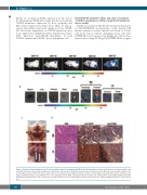
A
B
CD
Figure 6. Pattern of organ infiltration in the Toledo-Luci diffuse large B-cell lymphoma (DLBCL) disseminated mouse model. (A) Bioluminescent intensity (BLI) follow up by IVIS Spectrum of mice intravenously injected with Toledo cells transfected with the Luciferase gene (Toledo-Luci cells). (B) Ex vivo representative images of the recorded BLI emission in different mouse organs: spleen, liver, kidneys, heart, lungs, hind limbs, cranium, cervical lymph nodes (LN) and renal LN. (C) Macroscopic images showing Toledo-Luci cell infiltration in cervical LN and renal LN. White arrows show the LN location. (D) Hematoxylin & Eosin (H&E) staining, anti-CD20 immunohistochemistry (IHC) for B-cell detection, and anti-CXCR4 IHC in bone marrow (BM) (cranium) and LN (cervical). Original magnification x400. Scale bars=50 mm.
748
haematologica | 2020; 105(3)
