
Page 233 - Haematologica March 2020
P. 233
CXCR4-targeted nanocarrier to DLBCL cells
staining co-localized (yellow) on the cell membrane. Moreover, similar to findings in SC Toledo tumors, in the disseminated model, we found a release of the nanocarrier into CXCR4+ DLBCL cell cytosol separated from endocyt- icvesiclescontainingtheCXCR4receptor(Figure7C).
T22-GFP-H6 internalization in CXCR4+ mouse cells
To support the relevance of our CXCR4+ DLBCL models for clinical translation of the tumor (human cells) and non- tumor (mouse cells) biodistribution data, we assessed whether the nanocarrier internalized in mouse cells.
E
AB
C
D
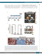
Figure 5. CXCR4-dependent uptake of T22-GFP-H6 nanocarrier in diffuse large B-cell lymphoma (DLBCL) subcutaneous (SC) tumors. (A) For the CXCR4 blocking experiment, mice were injected with a total of three SC doses of AMD3100 at 10 mg/kg. The time point of mice sacrifice was 2 hours (h) after the last AMD3100 SC injection, which corresponds to the 5h fluorescence intensity (FLI) peak after T22-GFP-H6 injection. (B) Representative images of emitted FLI by SC Toledo tumors from buffer, T22-GFP-H6, AMD3100 or AMD3100+T22-GFP-H6 treated animals. (C) FLI levels of SC tumor-bearing-mice of Toledo cells administered with T22-GFP- H6 or AMD3100+T22-GFP-H6 and FLI levels of the tumors in T22-GFP-H6-treated bearing SC CXCR4+ SUDHL-2 tumors or SC CXCR4– SUDHL-2 tumors. (D) A repre- sentative image of the FLI in SC tumors of CXCR4– SUDHL-2 and CXCR4+ SUDHL-2 cells after 5h of T22-GFP-H6 or buffer administration. (E) Level of membrane CXCR4 expression detected by immunohistochemistry (IHC) in SC tumors derived from Toledo, CXCR4+ SUDHL-2 and CXCR4– SUDHL-2 cells. **P<0.01. Original magnification x1000.
haematologica | 2020; 105(3)
747
