
Page 221 - Haematologica March 2020
P. 221
CDCA7 promotes lymphoma invasion
for ACTN1 barely detected its expression in control-trans- duced cells (Figure 6A-D). However, ACTN1 was readily detected in CDCA7-knockdown cells (Figure 6A-D). In contrast, a polyclonal antibody that reacts with isoforms 1, 2, and 4 showed a similar staining intensity in control and silenced cells (Figure 6A-D). These results suggested that CDCA7 might inhibit ACTN1 expression. However, immunoblot analysis of these cells with the monoclonal antibody revealed similar ACTN1 levels in control and silenced cells (Figure 6E). Together, these data suggest that CDCA7 silencing unmasks the epitope recognized by the ACTN1-specific monoclonal antibody.
transduced lymphoma cells and its polarized distribution markedly decreased upon CDCA7 silencing (Figure 7A- D). Of note, this redistribution was accompanied by a substantial increase of pMLC-S19 levels in silenced cells (Figure 7A, B, E). MLC phosphorylation can be induced by RhoA kinase (ROCK).33 To determine whether ROCK- mediated MLC activation contributed to the inhibition of cell migration imposed by CDCA7 knockdown, we treat- ed control and lymphoma cells with the ROCK inhibitor fasudil. We found that fasudil inhibited MLC phosphory- lation (Online Supplementary Figure S8) and neutralized the inhibition of cell migration in CDCA7-silenced lymphoma cells (Figure 8). Similarly, the NM-II inhibitor blebbistatin restored the migration competency of CDCA7-silenced cells (Figure 8), suggesting that ROCK-mediated NM-II activation hindered cell migration upon CDCA7 knock- down. Given the increase of F-actin in CDCA7-silenced cells, we also investigated the contribution of actin poly- merization to the inhibition of lymphoma cell migration. We found that treatment of these cells with the actin poly- merization inhibitor cytochalasin D overcame the migra- tory restraint imposed by CDCA7 silencing (Figure 8). Together, these results strongly support the notion that CDCA7 modulation of myosin activation and actin poly- merization is critical for the regulation of cell migration.
Discussion
While the processes and mechanisms involved in carci- noma invasion and the formation of metastases have been extensively characterized, little is known about the molec- ular mechanisms involved in lymphoma cell invasion. Here we show that CDCA7 is a critical mediator of lym- phoma cell invasion in vivo and in vitro and that CDCA7 knockdown greatly impairs lymphoma migration, through the regulation of tubulin and actomyosin cytoskeleton dynamics.
Metastases involve breaching of numerous histological barriers to move to distant sites. In the case of epithelial cancers, this process involves not only cell motility but also the proteolytic degradation of ECM molecular com- ponents. Among hundreds of proteinase genes, the MMP family has been implicated in carcinoma tumor invasion and metastasis formation.4 Indeed, MMP are overex-
Staining of α-actinin with the polyclonal antibody showed a dotted pattern in control-transduced BL-2 and Toledo cells and, contrary to the actin and tubulin cytoskeletons, its distribution was not substantially affect- ed by CDCA7 silencing (Online Supplementary Figure S7A, B). As one of the roles of α-actinin is to act as a link between integrins and the actin cytoskeleton, we investi- gated the distribution of active β1 integrins in lymphoma cells. Similar to α-actinin, active β1 integrin staining showed a dotted pattern in control- and CDCA7-knock- down BL-2 and Toledo cells (Online Supplementary Figure S7A, B). The presence of numerous white dots in merged images (Online Supplementary Figure S7A, B) strongly sug- gested that α-actinin and active β1 integrins do indeed colocalize in these cells. Determination of Pearson and Mander coefficients supported this hypothesis (Online Supplementary Figure S7C, D).
The actomyosin cytoskeleton is constituted by F-actin in association with numerous proteins, including myosins and tropomyosins (TPM). To investigate whether CDCA7 also regulates the cellular distribution of these proteins, we used fluorescence microscopy analysis. We found that TPM3 showed a polarized distribution in nearly 60% of control-transduced BL-2 and Toledo lymphoma cells, which was markedly decreased upon CDCA7 knock- down (Figure 7A-D).
As phosphorylation of the myosin regulatory light chain (MLC) on Ser19 is a marker of NM-II activation,11 we investigated the distribution of active myosin in lym- phoma cells by immunofluorescence using an antibody that specifically recognizes MLC phosphorylated on that residue (pMLC-S19). Similar to TPM3 and F-actin, pMLC- S19 was located in one pole of nearly 40% of control-
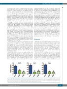
Figure 4. CDCA7 knockdown markedly inhibits serum-induced lymphoma migration. DG-75, BL-2, and Toledo cells were transduced with lentivirus encoding the indicated short hairpin (sh) RNA and seeded on the upper surface of the fibronectin-coated polycarbonate membrane of transwell chambers containing 10% fetal bovine serum in the lower chamber. Quantification of the relative migration capacity is shown as the mean + standard error of mean of three independent transduc- tions. *P<0.05, **P<0.01 and ***P<0.001 (one-way analysis of variance with the Bonferroni post-test).
haematologica | 2020; 105(3)
735
