
Page 219 - Haematologica March 2020
P. 219
CDCA7 promotes lymphoma invasion
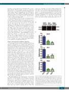
than that of control-transduced cells (Figure 2B). To deter- mine whether CDCA7 mediates invasion of other lym- phoma cells, we transduced BL-2 (Burkitt lymphoma) and Toledo (diffuse large B-cell lymphoma) cells with sh-25 or sh-83 lentivirus. These shRNA readily silenced CDCA7 expression in these cells (Figure 2A, middle and right pan- els) and sharply decreased their invasive capacity relative to that of control cells (Figure 2B).
As the zebrafish is a robust model for studying the inva- sive behavior of human tumor cells,29 we used it to evalu- ate the contribution of CDCA7 to lymphoma invasion in vivo. Transduced DG-75 cells were stained with live dye DiI and microinjected into the yolk sac of zebrafish embryos. The capacity of these cells to escape the yolk sac and migrate to the embryo tail was quantified as the per- cent of embryos with >5 labeled lymphoma cells in the tail. While control cells were found in the tail of nearly 60% of the embryos, less than 40% of the embryos inoc- ulated with CDCA7-silenced cells showed lymphoma cells in the tail (Figure 3A, B). To assess whether these results could be extended to other lymphomas, we deter- mined the capacity of control and CDCA7-silenced Toledo cells to migrate from the yolk sac to the embryo tail. We found that CDCA7 knockdown markedly decreased the presence of Toledo cells in the tail (Figure 3C, D). Together, our results strongly suggest that CDCA7 is a key mediator of lymphoma invasion.
CDCA7 silencing hinders lymphoma migration
To investigate the mechanisms underlying CDCA7 reg- ulation of cell invasion, we analyzed the expression of MMP2 and MMP9, the major metalloproteinases involved in basement membrane and stromal ECM degradation during invasion.5 The expression of these metallopro- teinases was not detected in DG-75, BL-2, and Toledo cells (Online Supplementary Figure S2A), but it was readily detected in breast cancer MCF-7 or colon carcinoma SW480 cells (Online Supplementary Figure S2B). Since these results suggest that ECM degradation is not required for lymphoma invasion, we hypothesized that the migratory capacity of lymphoma cells might be critical for invasion. We therefore assessed the contribution of CDCA7 to lym- phoma cell migration using fibronectin-coated transwell plates and FBS as a chemoattractant stimulus. BL-2 and Toledo cells attached poorly to fibronectin, but their bind- ing was stimulated in the presence of the TS2/16 mono- clonal antibody (Online Supplementary Figure S3A), an anti- integrin β1 monoclonal antibody that increases the avidity and affinity of β1 integrins for their ligands.30 Of note, we could not detect adhesion of DG-75 cells to fibronectin even in the presence of this antibody (not shown). As lym- phoma cells bind poorly to fibronectin, they reach the lower transwell chamber instead of remaining attached to the fibronectin-coated filter. Quantification of the number of cells in the lower chamber showed that CDCA7 silenc- ing markedly decreased the migratory capacity of DG-75, BL2, and Toledo cells (Figure 4).
Although lymphoma cells bind poorly to fibronectin, the ablation of this binding could formally account for the inhibition of cell migration upon CDCA7 silencing. Alternatively, a sharp increase in binding could also slow down migration. However, we found that CDCA7 knock- down did not substantially affect the binding of lym- phoma cells to fibronectin (Online Supplementary Figure S3A). Moreover, CDCA7 silencing did not affect the
expression of integrins α4 and β1 (Online Supplementary Figure S3B, C), the subunits of the major fibronectin recep- tor of these cells. Activation of β1 integrin binding activity induces a conformational modification of the β1 subunit that is recognized by the HUTS-21 monoclonal antibody.31 Staining of lymphoma cells with HUTS-21 showed that CDCA7 silencing did not affect the activity of this fibronectin receptor (Online Supplementary Figure S3B, C).
A
B
Figure 2. CDCA7 knockdown inhibits lymphoma invasion in vitro. (A) Representative CDCA7 and α-tubulin immunoblot analysis of cell lysates from DG-75, BL-2, and Toledo cells lentivirally transduced with the indicated short hairpin (sh) RNA. (B) These cells were seeded on the upper surface of the matrigel-coated polycarbonate membrane of transwell chambers containing 10% fetal bovine serum in the lower chamber. Quantification of the relative inva- sive capacity is shown as the mean + standard error of mean. of three independ- ent transductions. *P<0.05 and **P<0.01 (one-way analysis of variance with the Bonferroni post-test).
haematologica | 2020; 105(3)
733
