
Page 218 - Haematologica March 2020
P. 218
C. Martín-Cortázar et al.
Online Supplementary Table S1). These non-lymphoid tis- sues were extremely disorganized and embedded within the tumor (Figure 1B, C and Online Supplementary Table S1), suggesting that lymphoma cells invaded the neighbor- ing fat or muscle. In contrast, only 40% of tumors formed by CDCA7-silenced cells contained non-tumoral tissues and, when present, these tissues showed a rather well- preserved organization (Figure 1B, C and Online Supplementary Table S1). These results therefore suggest that while control lymphoma cells readily invade and dis- organize adjacent tissues, CDCA7-silenced lymphoma cells hardly invade them. We looked for gene expression profiles of metastatic lymphomas using Genevestigator.28 While we found gene expression profiling data of more than 1,600 cases of lymphoid tumors, we only found data on four metastatic cases (Online Supplementary Figure S1). Of note, CDCA7 levels were high in these cases and in numerous non-metastatic lymphoma/leukemia samples
AC
(Online Supplementary Figure S1), suggesting that CDCA7 might be clinically relevant.
CDCA7 silencing restrains lymphoma invasion in vitro and in vivo
To confirm the contribution of CDCA7 to lymphoma cell invasion, we determined the capacity of CDCA7- silenced cells to invade matrigel-coated transwell plates. CDCA7 knockdown in DG-75 cells transduced with lentivirus encoding sh-Ctl, sh-25 or sh-83 was confirmed by immunoblotting (Figure 2A, left panel). Transduced cells were suspended in serum-free medium and seeded in the top chamber of matrigel-coated transwell plates. We used fetal bovine serum (FBS) as a chemoattractant in the lower chamber of these plates. Quantification of the num- ber of cells capable of crossing the matrigel barrier and reaching the lower chamber showed that the invasive capacity of CDCA7-silenced cells was markedly lower
B
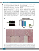
Figure 1. CDCA7 silencing limits lymphoma dissociation of neighboring tissues. DG-75 cells were transduced with lentivirus encoding short hairpin (sh) control (Ctl) RNA or the CDCA7-specific shRNA, sh-25 and sh-83, and selected in the presence of puromycin for >5 days. (A) Representative CDCA7 and α-tubulin (loading control) immunoblot analysis of these cells. (B, C) Transduced DG-75 cells were inoculated subcutaneously in immunodeficient NOD-SCID mice and tumors grown after 3 weeks were embedded in paraffin. All mice were inoculated with control cells in one flank (n=13) and sh-25 (n=7) or sh-83 (n=6) cells in the opposite flank. (B) Representative images of tumor sections from indicated mice stained with hematoxylin. Massively infiltrated (top panels) and poorly or non-infiltrated muscle and fat tissues (bottom panels) are shown. (C) Percentage of tumor masses with presence of non-tumoral tissues (NT) and percentage of tumor masses with heavily infil- trated non-tumoral tissues (HI). Additional information on these tumors is shown in Online Supplementary Table S1.
732
haematologica | 2020; 105(3)
