
Page 102 - Haematologica March 2020
P. 102
A. Caulier et al.
UO126 induced spontaneous GPA expression in the absence of EPO, showing that ERK activation was neces- sary to maintain UT7/GM cells in an undifferentiated GPAlow state. YODA1 did not revert the high GPA expres- sion induced by ERK1/2 inactivation (Figure 6D). The same results were observed using a retrovirus containing a MEK dominant-negative form (Online Supplementary Figure S7B). This indicated that the effect of YODA1 required a functional ERK pathway. Phospho-Flow experiments showed that YODA1 induced strong ERK phosphoryla- tion in UT7/GM cells (Figure 6E). This was confirmed by western blot analysis (data not shown). ERK phosphoryla- tion was Ca2+-dependent, since it decreased strongly in the presence of EGTA (Figure 6E), and PIEZO1-dependent, since it was abrogated in cells transduced with Sh-PIEZO1 lentivirus (Figure 6F). In human primary erythroid progen- itors, no p-ERK1/2 was detected in the absence of EPO. In the presence of EPO, YODA1 synergized with EPO for ERK phosphorylation, consistent with a role for PIEZO1 in the modulation of EPO-dependent ERK signaling in pri- mary cells (Figure 6G). This effect was confirmed by west- ern blot analysis (Online Supplementary Figure S7C). We
AB
observed a similar synergistic effect in EPO-induced STAT5 phosphorylation in primary erythroid cells (Figure 6H), also confirmed by western blotting (Online Supplementary Figure S7D). Of note, no effect of YODA1 on STAT5 phosphorylation was seen in UT7/GM cells, in which STAT5-phopshorylation is not EPO-dependent (Online Supplementary Figure S7E). Taken together, these data argue for a role of PIEZO1 in modulating ERK and STAT5 signaling pathways downstream of EPO-receptor activation in human progenitor cells.
PIEZO1 gain-of-function mutations in hereditary xero- cytosis delay erythroid differentiation and mimic the effect of chemical activation
Since most HX patients have PIEZO1 gain-of-function mutations, we tested whether the same phenotype could be observed during in vitro erythroid differentiation from PIEZO1-HX progenitors. Fourteen patients from 11 fami- lies (HX #1 to #11) carrying ten different PIEZO1 muta- tions were tested (HX#3: 2 siblings; HX#10: 3 siblings). The patients’ characteristics are shown in Table 1. Nine phlebotomy samples from five patients [3 families, HX#1
C
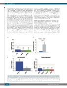
Figure 4. Effect of PIEZO1 activation on the transcriptional program of erythroid differentiation in primary human CD34+-derived cells, assessed by quantitative reverse transcriptase polymerase chain reaction. For all experiments, primary cells were cultured for 10 days, with 1 mM YODA1 or dimethylsulfoxide (DMSO) stim- ulation from day 3 to 10. Gene expression was assessed relative to GAPDH expression. (A) Compared to exposure to DMSO, exposure to 1 mM YODA1 decreased GPA mRNA expression (x0.49±0.17), β-globin RNA expression (x0.4±0.26) and α-globin RNA expression (x0.3±0.24). (B) Compared to exposure to DMSO, exposure to 1 mM YODA1 increased the GATA2/GATA1 mRNA ratio (x6.2±1.9). (C) Stimulation with 1 mM YODA1 increased STAT5A and BMI-1 expression, and decreased EPOR, SLC4A1, ALAS2, and AHSP mRNA expression. (n=3 for all experiments); ***P<0.001; **P<0.01; *P<0.05.
616
haematologica | 2020; 105(3)
